Copyright
©The Author(s) 2015.
World J Gastroenterol. May 28, 2015; 21(20): 6409-6416
Published online May 28, 2015. doi: 10.3748/wjg.v21.i20.6409
Published online May 28, 2015. doi: 10.3748/wjg.v21.i20.6409
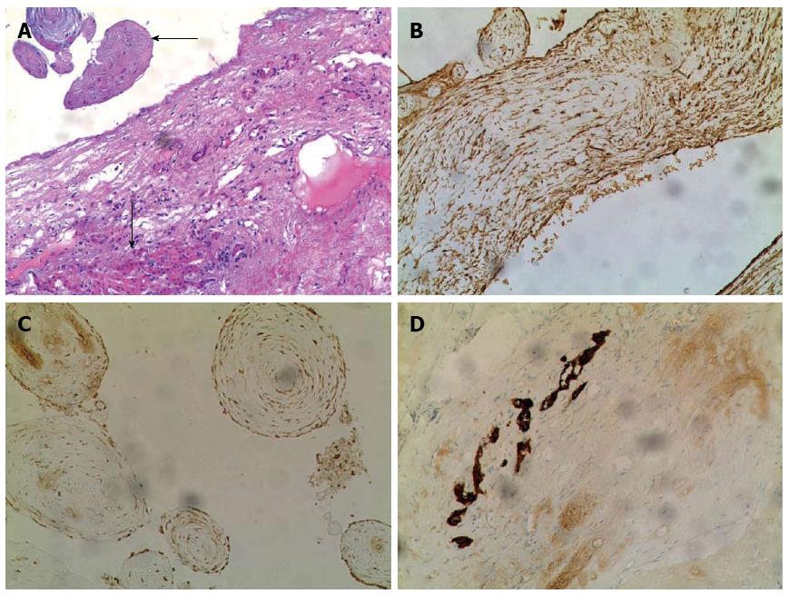
Figure 4 Clear boundary between liver parenchyma and proliferating connective tissue.
A: The mass consisted of loose connective tissue full of myxoid matrix forming visible cysts (upper arrow). Small amounts of remaining liver tissue, with a lack of lobular architecture, were located in peripheral areas (lower arrow) (HE, original magnification × 100); Myxoid stroma with spindle cells showing smooth muscle differentiation were confirmed by positive staining for vimentin (B) and smooth muscle actin (C) (original magnification × 100); Benign dilated bile ducts were confirmed by positive staining for cytokeratin 7 (D) (original magnification × 100).
- Citation: Li J, Cai JZ, Guo QJ, Li JJ, Sun XY, Hu ZD, Cooper DK, Shen ZY. Liver transplantation for a giant mesenchymal hamartoma of the liver in an adult: Case report and review of the literature. World J Gastroenterol 2015; 21(20): 6409-6416
- URL: https://www.wjgnet.com/1007-9327/full/v21/i20/6409.htm
- DOI: https://dx.doi.org/10.3748/wjg.v21.i20.6409


