Copyright
©The Author(s) 2015.
World J Gastroenterol. May 28, 2015; 21(20): 6409-6416
Published online May 28, 2015. doi: 10.3748/wjg.v21.i20.6409
Published online May 28, 2015. doi: 10.3748/wjg.v21.i20.6409
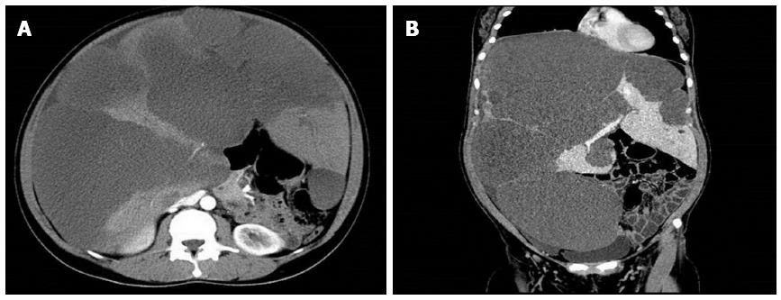
Figure 1 Contrast enhanced computed tomography of the abdomen revealed the near replacement of the liver with diffuse cystic masses of low density.
A: Enhanced computed tomography scan shows the near replacement of the liver with diffuse cystic masses, leaving only small amounts of liver parenchyma. The portal vein and inferior vena cava are obviously compressed; B: The massively enlarged liver essentially occupies the entire abdominal cavity, with other abdominal organs being compressed and displaced.
- Citation: Li J, Cai JZ, Guo QJ, Li JJ, Sun XY, Hu ZD, Cooper DK, Shen ZY. Liver transplantation for a giant mesenchymal hamartoma of the liver in an adult: Case report and review of the literature. World J Gastroenterol 2015; 21(20): 6409-6416
- URL: https://www.wjgnet.com/1007-9327/full/v21/i20/6409.htm
- DOI: https://dx.doi.org/10.3748/wjg.v21.i20.6409


