Copyright
©The Author(s) 2015.
World J Gastroenterol. May 28, 2015; 21(20): 6404-6408
Published online May 28, 2015. doi: 10.3748/wjg.v21.i20.6404
Published online May 28, 2015. doi: 10.3748/wjg.v21.i20.6404
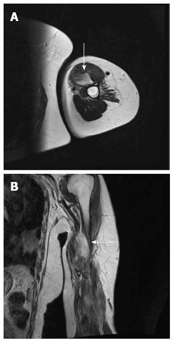
Figure 1 Magnetic resonance imaging of the left arm.
A: Axial T2-weighted image showing a huge, ill-defined, high-signal-intensity mass (solid arrow); B: Coronal T2-weighted image showing a high-signal-intensity mass (dotted arrow).
- Citation: Jin SS, Jeong HS, Noh HJ, Choi WH, Choi SH, Won KY, Kim DP, Park JC, Joung MK, Kim JG, Sul HJ, Lee SW. Gastrointestinal stromal tumor solitary distant recurrence in the left brachialis muscle. World J Gastroenterol 2015; 21(20): 6404-6408
- URL: https://www.wjgnet.com/1007-9327/full/v21/i20/6404.htm
- DOI: https://dx.doi.org/10.3748/wjg.v21.i20.6404


