Copyright
©The Author(s) 2015.
World J Gastroenterol. May 28, 2015; 21(20): 6398-6403
Published online May 28, 2015. doi: 10.3748/wjg.v21.i20.6398
Published online May 28, 2015. doi: 10.3748/wjg.v21.i20.6398
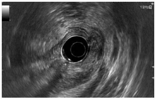
Figure 3 Endosonographic views.
A lobulated mass measuring about 5 cm with mixed echogenicity and an irregular margin was observed at the gastric antrum.
- Citation: Kim SB, Oh MJ, Lee SH. Gastric subepithelial lesion complicated with abscess: Case report and literature review. World J Gastroenterol 2015; 21(20): 6398-6403
- URL: https://www.wjgnet.com/1007-9327/full/v21/i20/6398.htm
- DOI: https://dx.doi.org/10.3748/wjg.v21.i20.6398


