Copyright
©The Author(s) 2015.
World J Gastroenterol. May 28, 2015; 21(20): 6391-6397
Published online May 28, 2015. doi: 10.3748/wjg.v21.i20.6391
Published online May 28, 2015. doi: 10.3748/wjg.v21.i20.6391
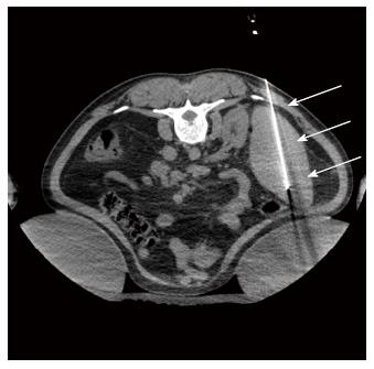
Figure 2 Axial non-enhanced computed tomography scan guiding the splenic radiofrequency ablation, with the patient in the prone position.
Note the “single” probe (arrow) in the middle/ lower third of the spleen. where the ablation zones were concentrated.
- Citation: Martins GLP, Bernardes JPG, Rovella MS, Andrade RG, Viana PCC, Herman P, Cerri GG, Menezes MR. Radiofrequency ablation for treatment of hypersplenism: A feasible therapeutic option. World J Gastroenterol 2015; 21(20): 6391-6397
- URL: https://www.wjgnet.com/1007-9327/full/v21/i20/6391.htm
- DOI: https://dx.doi.org/10.3748/wjg.v21.i20.6391


