Copyright
©The Author(s) 2015.
World J Gastroenterol. May 28, 2015; 21(20): 6384-6390
Published online May 28, 2015. doi: 10.3748/wjg.v21.i20.6384
Published online May 28, 2015. doi: 10.3748/wjg.v21.i20.6384
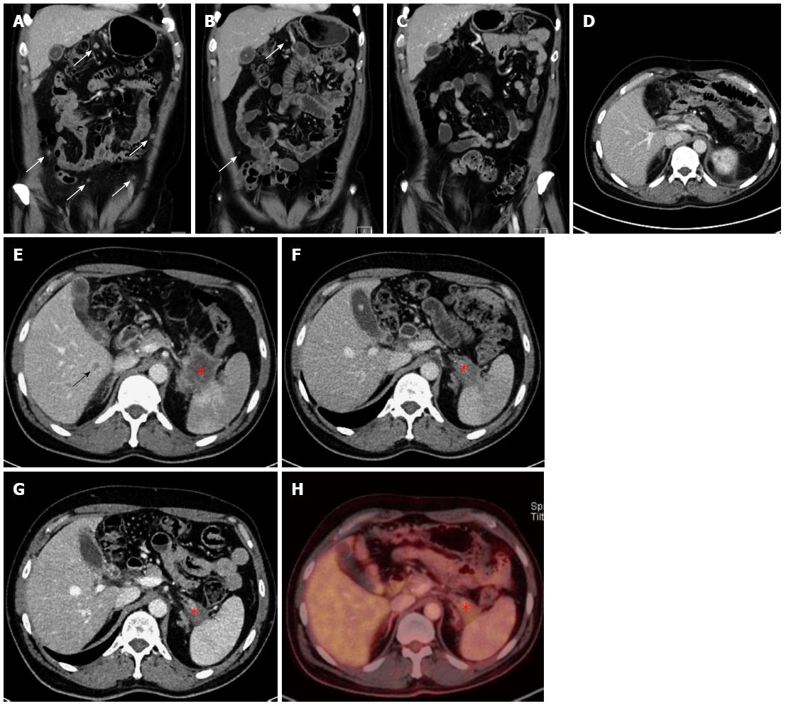
Figure 3 Computed tomography and positron emission tomography images of patient 2.
Coronal (A, B, C) contrast-enhanced CT images demonstrated complete regression of multiple peritoneal metastases (white arrow). Axial CT images (E, F, G) and PET-CT image (H) show a regredient mass (star) in the tail of the pancreas. Liver metastasis (black arrow) was seen only in the initial CT-examination (E). Post-operative CT (D) showed complete resection of the primary tumor in the pancreatic tail and no visible metastases. CT: Computed tomography; PET: Positron emission tomography.
- Citation: Schneitler S, Kröpil P, Riemer J, Antoch G, Knoefel WT, Häussinger D, Graf D. Metastasized pancreatic carcinoma with neoadjuvant FOLFIRINOX therapy and R0 resection. World J Gastroenterol 2015; 21(20): 6384-6390
- URL: https://www.wjgnet.com/1007-9327/full/v21/i20/6384.htm
- DOI: https://dx.doi.org/10.3748/wjg.v21.i20.6384


