Copyright
©The Author(s) 2015.
World J Gastroenterol. May 28, 2015; 21(20): 6384-6390
Published online May 28, 2015. doi: 10.3748/wjg.v21.i20.6384
Published online May 28, 2015. doi: 10.3748/wjg.v21.i20.6384
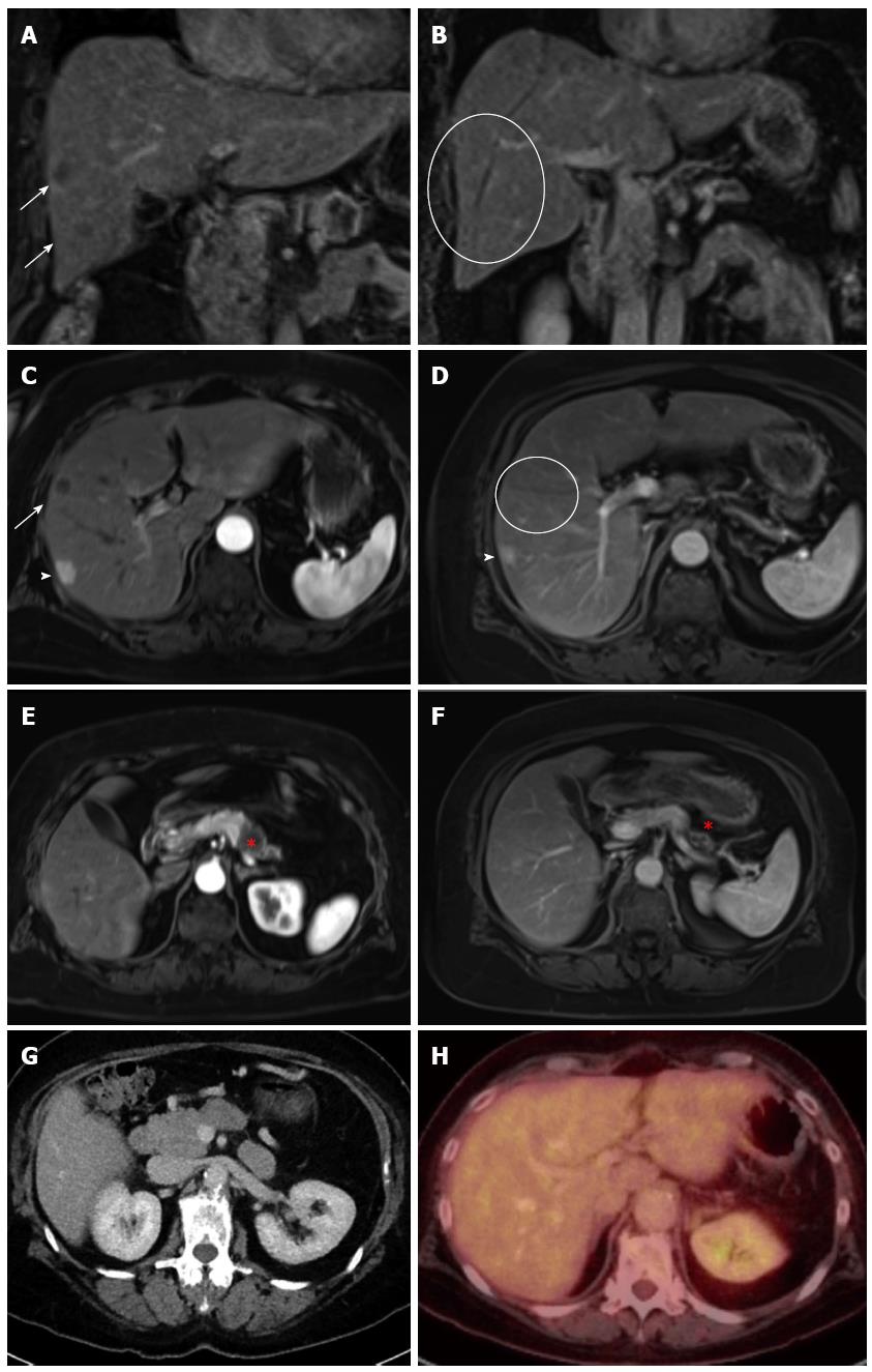
Figure 1 Computed tomography, magnetic resonance imaging and positron emission tomography images of patient 1.
Coronal (A, B) and transversal (C-F) T1w contrast-enhanced MRI images demonstrated regredient primary malignancy (star) in the tail of pancreas (E, F) and regredient hepatic metastases (arrow, cycle) in the right liver lobe (A, B, C, D). An additional hemangioma (arrowhead) was observed. Postoperative CT (G) and PET-CT (H) showed complete resection of the primary tumor and no visible metastasis. CT: Computed tomography; MRI: Magnetic resonance imaging; PET: Positron emission tomography.
- Citation: Schneitler S, Kröpil P, Riemer J, Antoch G, Knoefel WT, Häussinger D, Graf D. Metastasized pancreatic carcinoma with neoadjuvant FOLFIRINOX therapy and R0 resection. World J Gastroenterol 2015; 21(20): 6384-6390
- URL: https://www.wjgnet.com/1007-9327/full/v21/i20/6384.htm
- DOI: https://dx.doi.org/10.3748/wjg.v21.i20.6384


