Copyright
©The Author(s) 2015.
World J Gastroenterol. May 28, 2015; 21(20): 6180-6193
Published online May 28, 2015. doi: 10.3748/wjg.v21.i20.6180
Published online May 28, 2015. doi: 10.3748/wjg.v21.i20.6180
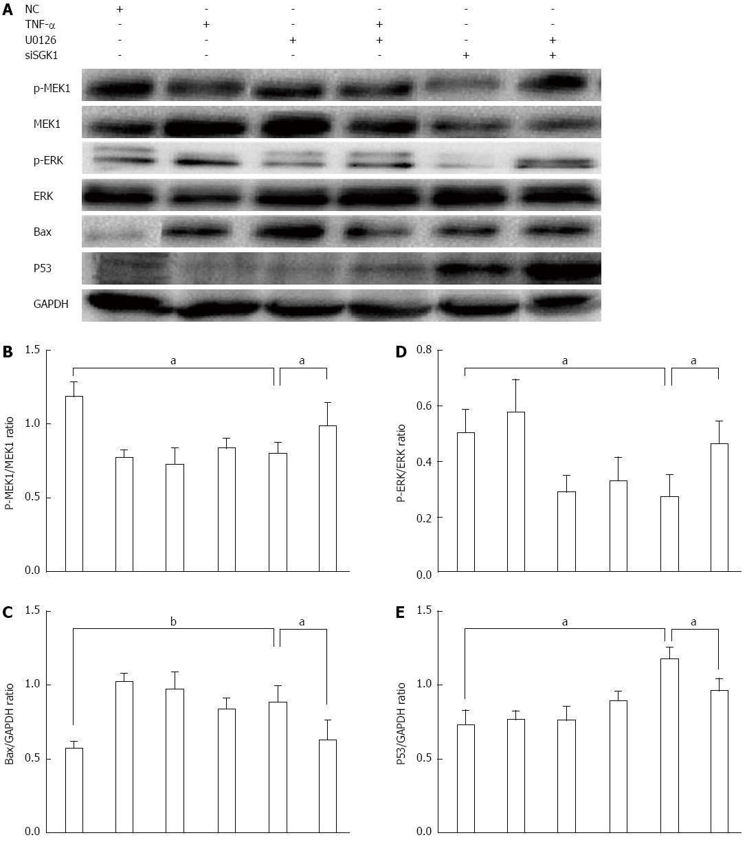
Figure 7 P53 and Bax are downstream of the mitogen-activated protein kinase kinase 1/extracellular signal regulated protein kinase pathway.
A: IECs in culture were pretreated with the MEK1 inhibitor (U0126) followed by stimulation with TNF-α and NC or SGK1 siRNA. Protein expression levels were then detected by western blotting; B: The level of MEK1 phosphorylation was assessed by the ratio of p-MEK1/MEK1; D: The level of ERK1/2 phosphorylation was assessed by the ratio of p-ERK/ERK; C, E: p53 and Bax expression levels were assessed by the ratios of p53/GAPDH and Bax/GAPDH, respectively. aP < 0.05 and bP < 0.01, siSGK1-treated group vs negative controls or the U0126-treated group vs the non-U0126-treated group before siSGK1 transfection. SGK1: Serum-and-glucocorticoid-inducible-kinase-1; TNF: Tumor necrosis factor; IECs: Intestinal epithelial cells; MEK: Mitogen-activated protein kinase kinase 1; ERK: Extracellular signal regulated protein kinase; GAPDH: Glyceraldehyde-3-phosphate dehydrogenase.
-
Citation: Bai JA, Xu GF, Yan LJ, Zeng WW, Ji QQ, Wu JD, Tang QY. SGK1 inhibits cellular apoptosis and promotes proliferation
via the MEK/ERK/p53 pathway in colitis. World J Gastroenterol 2015; 21(20): 6180-6193 - URL: https://www.wjgnet.com/1007-9327/full/v21/i20/6180.htm
- DOI: https://dx.doi.org/10.3748/wjg.v21.i20.6180


