Copyright
©The Author(s) 2015.
World J Gastroenterol. May 28, 2015; 21(20): 6180-6193
Published online May 28, 2015. doi: 10.3748/wjg.v21.i20.6180
Published online May 28, 2015. doi: 10.3748/wjg.v21.i20.6180
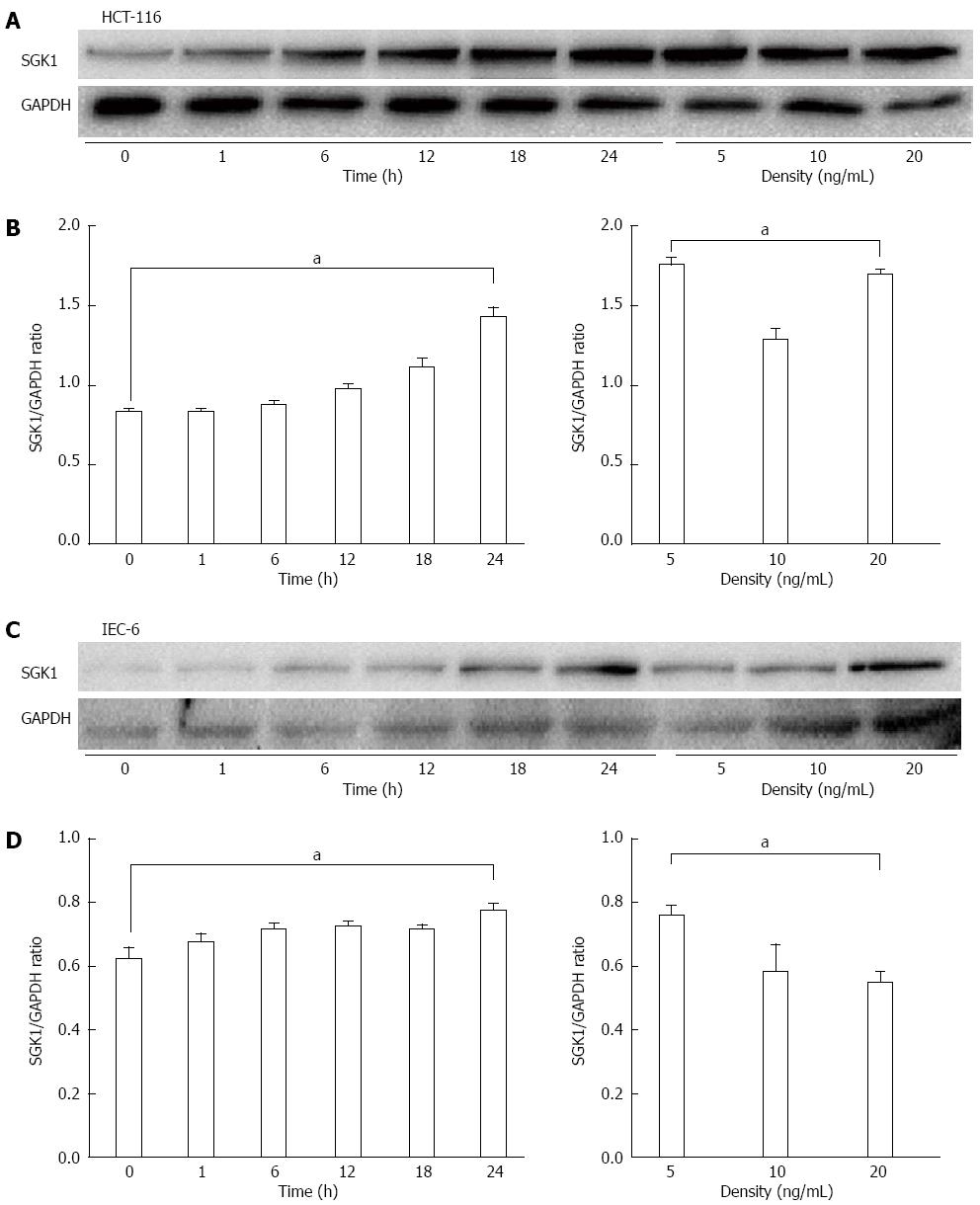
Figure 4 Serum-and-glucocorticoid-inducible-kinase-1 expression levels in tumor necrosis factor-treated intestinal epithelial cells assessed with western blotting.
A, B: SGK1 expression levels at different times and doses of TNF in HCT-116 cells; C, D: SGK1 expression levels at different times and doses of TNF in IEC-6 cells. aP < 0.05 and bP < 0.01 represented 24 h vs 0 h after TNF-treatment or 10 ng vs 5 ng TNF. GAPDH served as the internal control. SGK1: Serum-and-glucocorticoid-inducible-kinase-1; TNF: Tumor necrosis factor; IECs: Intestinal epithelial cells.
-
Citation: Bai JA, Xu GF, Yan LJ, Zeng WW, Ji QQ, Wu JD, Tang QY. SGK1 inhibits cellular apoptosis and promotes proliferation
via the MEK/ERK/p53 pathway in colitis. World J Gastroenterol 2015; 21(20): 6180-6193 - URL: https://www.wjgnet.com/1007-9327/full/v21/i20/6180.htm
- DOI: https://dx.doi.org/10.3748/wjg.v21.i20.6180


