Copyright
©The Author(s) 2015.
World J Gastroenterol. May 28, 2015; 21(20): 6180-6193
Published online May 28, 2015. doi: 10.3748/wjg.v21.i20.6180
Published online May 28, 2015. doi: 10.3748/wjg.v21.i20.6180
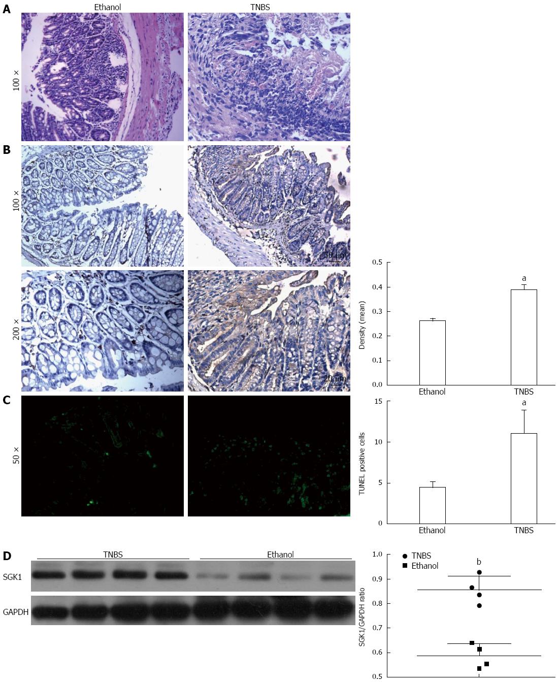
Figure 3 Serum-and-glucocorticoid-inducible-kinase-1 expression was increased in the colonic tissue of a 2,4,6-trinitrobenzene sulfonic acid-induced murine model.
A: Histological damage of colonic tissues induced by ethanol or 50% TNBS in mice (HE staining). Colonic tissues were obtained from the mice killed 3 d post-enema with ethanol or 50% TNBS; B: Immunohistochemical localization and upregulation of SGK1 expression in the colitis model. SGK1-dependent staining is indicated by brown areas. The density of positive areas was analyzed with Image Pro Plus; C: The TNBS group showed an increase in TUNEL positive cells. The positive cells were counted, and the results are presented as mean ± SD from six sample fields; D: Western blot analysis was performed to study the SGK1 level in TNBS-induced colitis compared with ethanol-treated mice. The ethanol-treated group served as controls in these experiments. aP < 0.05 and bP < 0.01 vs the ethanol treatment group. GAPDH served as the internal control.
-
Citation: Bai JA, Xu GF, Yan LJ, Zeng WW, Ji QQ, Wu JD, Tang QY. SGK1 inhibits cellular apoptosis and promotes proliferation
via the MEK/ERK/p53 pathway in colitis. World J Gastroenterol 2015; 21(20): 6180-6193 - URL: https://www.wjgnet.com/1007-9327/full/v21/i20/6180.htm
- DOI: https://dx.doi.org/10.3748/wjg.v21.i20.6180


