Copyright
©The Author(s) 2015.
World J Gastroenterol. May 28, 2015; 21(20): 6157-6166
Published online May 28, 2015. doi: 10.3748/wjg.v21.i20.6157
Published online May 28, 2015. doi: 10.3748/wjg.v21.i20.6157
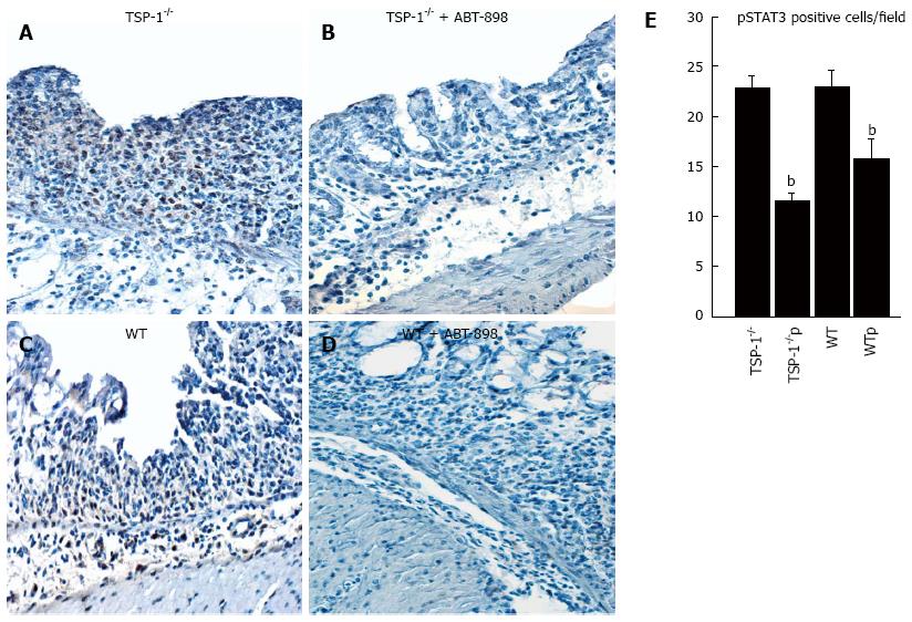
Figure 5 Activation of STAT3 by immunohistochemistry.
Immunohistochemistry for pSTAT3, in colitic tissues of TSP-1-/- controls (A), TSP-1-/- colonic tissues treated with ABT-898 (B), colons from WT mice (C) and from WT mice treated with the ABT-898 peptide (D), pSTAT3 positive brown staining was detected in the nucleus of epithelial cells and infiltrating leukocytes. Quantification of pSTAT3 positive cells was performed in colonic sections (E). Colons from WT and TSP-1-/- mice used as control showed higher numbers of positive cells when compared to mice treated with ABT-898. bP < 0.05 vs control. NS: Not significant; TSP-1-/-: TSP-1-/- controls; TSP-1-/-p: TSP-1-/- treated with ABT-898 peptide; WT: WT control; WTp: WT treated with ABT-898 peptide.
- Citation: Gutierrez LS, Ling J, Nye D, Papathomas K, Dickinson C. Thrombospondin peptide ABT-898 inhibits inflammation and angiogenesis in a colitis model. World J Gastroenterol 2015; 21(20): 6157-6166
- URL: https://www.wjgnet.com/1007-9327/full/v21/i20/6157.htm
- DOI: https://dx.doi.org/10.3748/wjg.v21.i20.6157


