Copyright
©The Author(s) 2015.
World J Gastroenterol. May 28, 2015; 21(20): 6157-6166
Published online May 28, 2015. doi: 10.3748/wjg.v21.i20.6157
Published online May 28, 2015. doi: 10.3748/wjg.v21.i20.6157
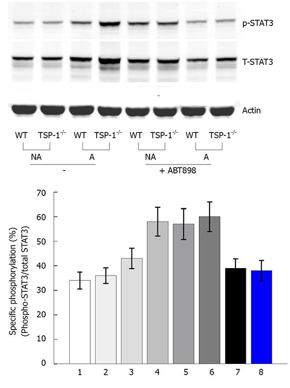
Figure 4 Activation of STAT3 by Western blotting.
Tissue lysates were prepared as described in “MATERIALS AND METHODS”, equal amounts of protein (80 μg) were resolved by 4%-12% SDS-PAGE, followed by Western blot with antibodies against STAT3, phosphorylated STAT3 (pSTAT3) (Ser727) and actin, respectively (bottom panel). STAT3 activation under acute colitis induced by DSS and the effects of the ABT-898 peptide on STAT3 activation during DSS-induced acute colitis; Top panel shows the specific phosphorylation of STAT3 from three repeated Western blots as represented by the bottom panel. The average values with standard deviations are showed. NA: Not affected colon tissue (no DSS); A: DSS induced colon tissue; TSP-1-/-: TSP-1-/- controls; TSP-1-/-p: TSP-1-/- treated with ABT-898 peptide; WT: WT control.
- Citation: Gutierrez LS, Ling J, Nye D, Papathomas K, Dickinson C. Thrombospondin peptide ABT-898 inhibits inflammation and angiogenesis in a colitis model. World J Gastroenterol 2015; 21(20): 6157-6166
- URL: https://www.wjgnet.com/1007-9327/full/v21/i20/6157.htm
- DOI: https://dx.doi.org/10.3748/wjg.v21.i20.6157


