Copyright
©The Author(s) 2015.
World J Gastroenterol. Jan 14, 2015; 21(2): 578-583
Published online Jan 14, 2015. doi: 10.3748/wjg.v21.i2.578
Published online Jan 14, 2015. doi: 10.3748/wjg.v21.i2.578
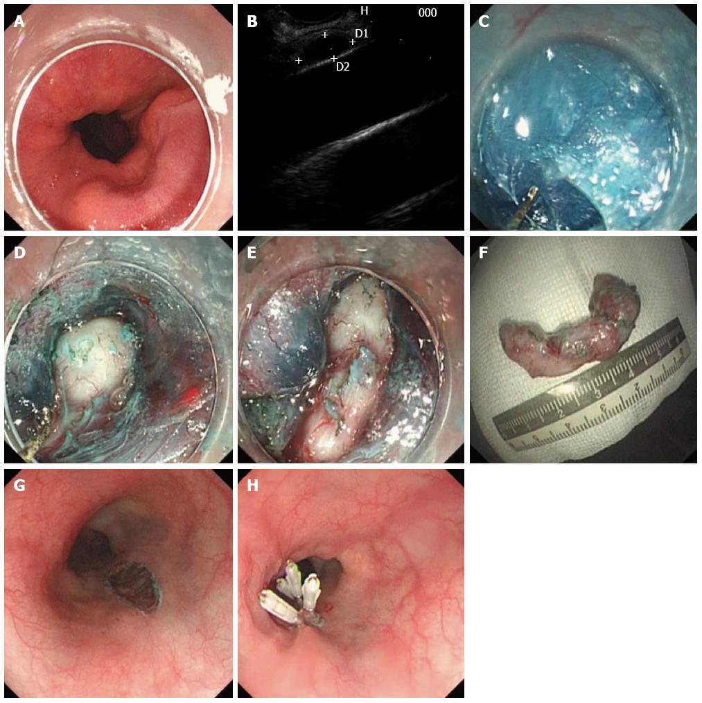
Figure 1 Submucosal tunneling and endoscopic resection of subepithelial tumors at the esophagogastric junction originating from the muscularis propria layer.
A: Endoscopic view of subepithelial tumors; B: Endoscopic ultrasonographic evaluation of the same lesion; C: Submucosal tunnel to the lesion (with a hook knife); D: The exposed tumor; E: Annular growth of the tumor; F: The resected specimen; G: The mucosal entry incision; H: The closure of the mucosal entry incision (with several clips).
- Citation: Zhou DJ, Dai ZB, Wells MM, Yu DL, Zhang J, Zhang L. Submucosal tunneling and endoscopic resection of submucosal tumors at the esophagogastric junction. World J Gastroenterol 2015; 21(2): 578-583
- URL: https://www.wjgnet.com/1007-9327/full/v21/i2/578.htm
- DOI: https://dx.doi.org/10.3748/wjg.v21.i2.578


