Copyright
©The Author(s) 2015.
World J Gastroenterol. Jan 14, 2015; 21(2): 432-438
Published online Jan 14, 2015. doi: 10.3748/wjg.v21.i2.432
Published online Jan 14, 2015. doi: 10.3748/wjg.v21.i2.432
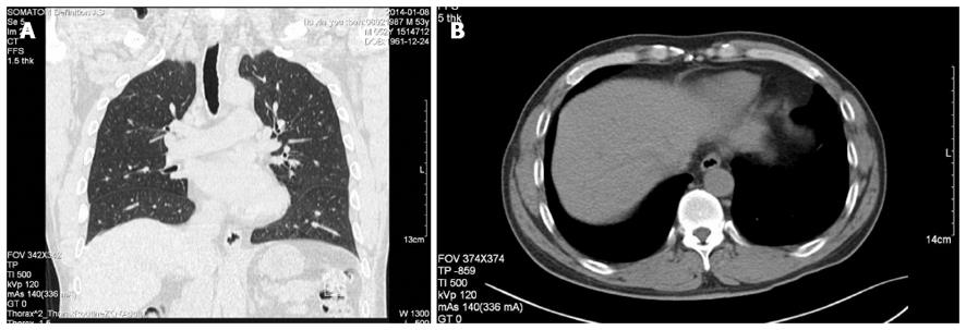
Figure 1 Computed tomography.
A: Frontal abdominal contrast-enhanced computed tomography (CT); B: Transversal abdominal CT demonstrating a homogeneous, low-density and well-circumscribed, subserosal cystic mass on the lesser curvature of the gastric cardia.
- Citation: Geng YH, Wang CX, Li JT, Chen QY, Li XZ, Pan H. Gastric foregut cystic developmental malformation: Case series and literature review. World J Gastroenterol 2015; 21(2): 432-438
- URL: https://www.wjgnet.com/1007-9327/full/v21/i2/432.htm
- DOI: https://dx.doi.org/10.3748/wjg.v21.i2.432


