Copyright
©The Author(s) 2015.
World J Gastroenterol. May 21, 2015; 21(19): 5867-5876
Published online May 21, 2015. doi: 10.3748/wjg.v21.i19.5867
Published online May 21, 2015. doi: 10.3748/wjg.v21.i19.5867
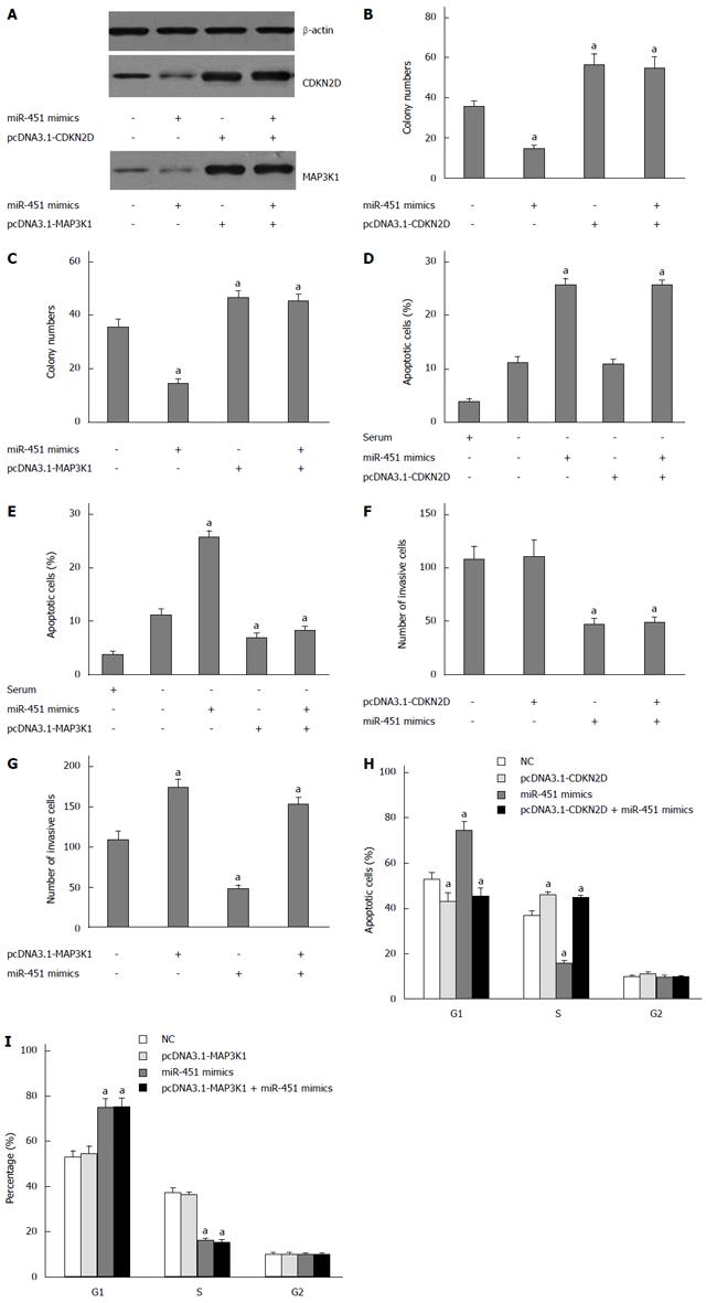
Figure 2 CDKN2D and MAP3K1 overexpression reverses the effect of miR-451.
A: CDKN2D and MAP3K1 protein levels were detected by Western blot assay. Western blot assay showed that transfection of miR-451 mimics inhibited the expression of CDKN2D or MAP3K1. Co-transfection of pcDNA3.1-CDKN2D or pcDNA3.1-MAP3K1and miR-451 abrogated the effects of miR-451 on CDKN2D or MAP3K1 expression. β-actin was used as a reference; B: The expression of CDKN2D could partially reverse the anti-proliferation function of miR-451. Colony formation assays were performed. aP < 0.05 vs control group; C: The expression of MAP3K1 could partially reverse the anti-proliferation function of miR-451. Colony formation assays were performed. aP < 0.05 vs control group; D: The expression of CDKN2D did not reverse the pro-apoptotic function of miR-451. Cells were transfected with pcDNA3.1-CDKN2D (not including 3’-UTR) or (and) miR-451. The cell apoptosis was assessed using flow cytometry assay; E: The expression of MAP3K1 reversed the pro-apoptotic function of miR-451. Cells were transfected with pcDNA3.1-MAP3K1 (not including 3’-UTR) or (and) miR-451. The cell apoptosis was assessed using flow cytometry assay; F: The expression of CDKN2D reversed the anti-migration function of miR-451. Cells were transfected with pcDNA3.1-CDKN2D (not including 3’-UTR) or (and) miR-451. The cell invasion was assessed using transwell assay; G: The expression of MAP3K1 reversed the anti-migration function of miR-451. Cells were transfected with pcDNA3.1-MAP3K1 (not including 3’-UTR) or (and) miR-451. The cell invasion was assessed using transwell assay; H: The expression of CDKN2D reversed G1 arrest of miR-451. Cells were transfected with pcDNA3.1-CDKN2D (not including 3’-UTR) or (and) miR-451. The cell cycle was assessed using flow cytometry assay; I: The expression of MAP3K1 did not reverse G1 arrest of miR-451. Cells were transfected with pcDNA3.1-MAP3K1 (not including 3’-UTR) or (and) miR-451. The cell cycle was assessed using flow cytometry assay (aP < 0.05 vs control group).
- Citation: Zang WQ, Yang X, Wang T, Wang YY, Du YW, Chen XN, Li M, Zhao GQ. MiR-451 inhibits proliferation of esophageal carcinoma cell line EC9706 by targeting CDKN2D and MAP3K1. World J Gastroenterol 2015; 21(19): 5867-5876
- URL: https://www.wjgnet.com/1007-9327/full/v21/i19/5867.htm
- DOI: https://dx.doi.org/10.3748/wjg.v21.i19.5867


