Copyright
©The Author(s) 2015.
World J Gastroenterol. May 21, 2015; 21(19): 5831-5842
Published online May 21, 2015. doi: 10.3748/wjg.v21.i19.5831
Published online May 21, 2015. doi: 10.3748/wjg.v21.i19.5831
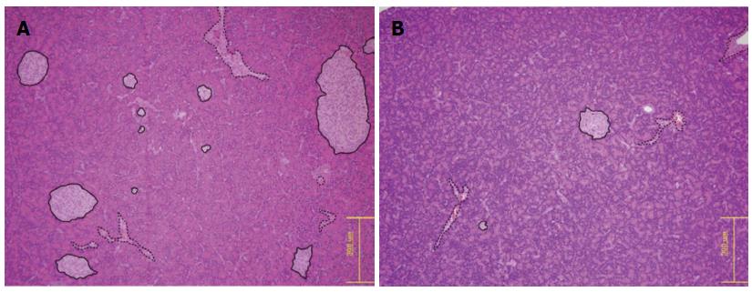
Figure 7 Hematoxylin and eosin staining of the pancreas of a control rat and a diabetic rat.
A light microscope at magnification × 100. All islets are outlined with solid lines, and the structures outlined with dotted line are the vessels. Both the islet size and the islet number were decreased in the diabetic rats (B) compared with the control rats (A).
- Citation: Cho HR, Lee Y, Doble P, Bishop D, Hare D, Kim YJ, Kim KG, Jung HS, Park KS, Choi SH, Moon WK. Magnetic resonance imaging of the pancreas in streptozotocin-induced diabetic rats: Gadofluorine P and Gd-DOTA. World J Gastroenterol 2015; 21(19): 5831-5842
- URL: https://www.wjgnet.com/1007-9327/full/v21/i19/5831.htm
- DOI: https://dx.doi.org/10.3748/wjg.v21.i19.5831


