Copyright
©The Author(s) 2015.
World J Gastroenterol. May 14, 2015; 21(18): 5739-5743
Published online May 14, 2015. doi: 10.3748/wjg.v21.i18.5739
Published online May 14, 2015. doi: 10.3748/wjg.v21.i18.5739
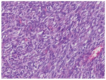
Figure 4 Histologic examination.
Malignant gastrointestinal neuroectodermal tumor consisting of spindle cells with eosinophilic and clear cytoplasm and vesicular nuclei was noted. (HE stain, × 200).
- Citation: Kim SB, Lee SH, Gu MJ. Esophageal subepithelial lesion diagnosed as malignant gastrointestinal neuroectodermal tumor. World J Gastroenterol 2015; 21(18): 5739-5743
- URL: https://www.wjgnet.com/1007-9327/full/v21/i18/5739.htm
- DOI: https://dx.doi.org/10.3748/wjg.v21.i18.5739


