Copyright
©The Author(s) 2015.
World J Gastroenterol. May 7, 2015; 21(17): 5432-5441
Published online May 7, 2015. doi: 10.3748/wjg.v21.i17.5432
Published online May 7, 2015. doi: 10.3748/wjg.v21.i17.5432
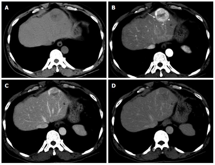
Figure 1 Computed tomography of the liver.
A: The entire mass was visualized as low density on non-contrast computed tomography (CT); B: Contrast-enhanced CT showed a strongly enhanced mass in the arterial phase, and the center of the mass was weakly enhanced; the internal component-like structure showed relatively strong enhancement with a blotchy vascular pattern within the tumor (arrowhead) and distorted vessels (arrow); C and D: The portal (C) and late (D) phase CT scans showed washout of the contrast medium, and a portion of the internal area was weakly enhanced.
- Citation: Maebayashi T, Abe K, Aizawa T, Sakaguchi M, Ishibashi N, Abe O, Takayama T, Nakayama H, Matsuoka S, Nirei K, Nakamura H, Ogawa M, Sugitani M. Improving recognition of hepatic perivascular epithelioid cell tumor: Case report and literature review. World J Gastroenterol 2015; 21(17): 5432-5441
- URL: https://www.wjgnet.com/1007-9327/full/v21/i17/5432.htm
- DOI: https://dx.doi.org/10.3748/wjg.v21.i17.5432


