Copyright
©The Author(s) 2015.
World J Gastroenterol. May 7, 2015; 21(17): 5259-5270
Published online May 7, 2015. doi: 10.3748/wjg.v21.i17.5259
Published online May 7, 2015. doi: 10.3748/wjg.v21.i17.5259
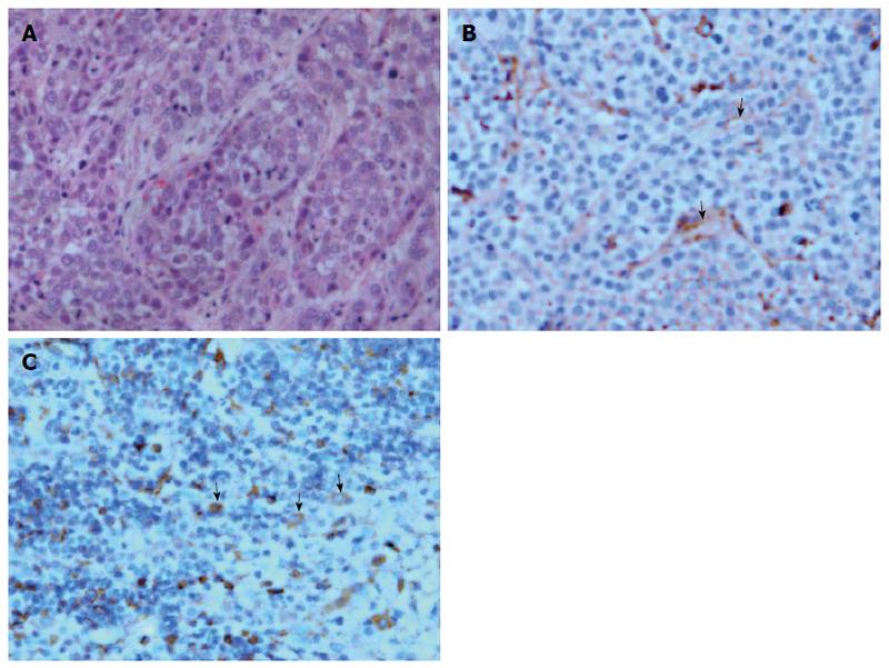
Figure 5 Histological and immunohistochemical micrographs of hepatic VX2 tumor.
A: Tumor tissue stained with hematoxylin and eosin (magnification × 400) demonstrates that the normal hepatic sinusoidal capillary disappeared and was replaced by a large amount of tumor cells; B: CD31 staining of tumor tissue shows the formation of new microvessels with incomplete layer of endothelial wall (arrows, magnification × 400); C: Vascular endothelial growth factor staining of tumor tissue depicts positive expression in the cytoplasm of tumor cell (arrows, magnification × 400).
- Citation: Zhang CY, Cui YF, Guo C, Cai J, Weng YF, Wang LJ, Wang DB. Low contrast medium and radiation dose for hepatic computed tomography perfusion of rabbit VX2 tumor. World J Gastroenterol 2015; 21(17): 5259-5270
- URL: https://www.wjgnet.com/1007-9327/full/v21/i17/5259.htm
- DOI: https://dx.doi.org/10.3748/wjg.v21.i17.5259


