Copyright
©The Author(s) 2015.
World J Gastroenterol. May 7, 2015; 21(17): 5250-5258
Published online May 7, 2015. doi: 10.3748/wjg.v21.i17.5250
Published online May 7, 2015. doi: 10.3748/wjg.v21.i17.5250
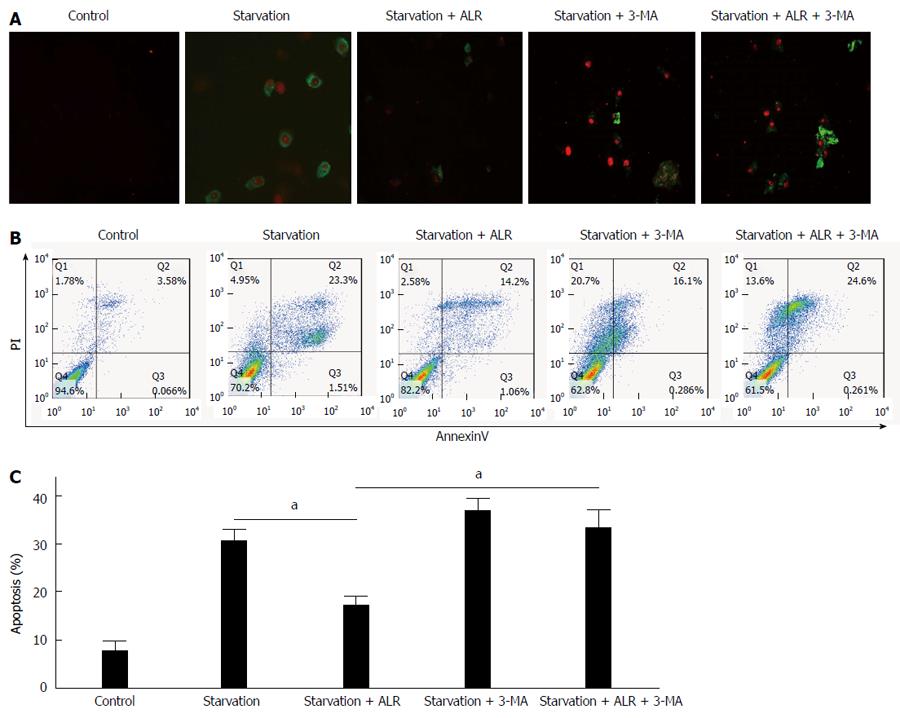
Figure 4 Role of autophagy in the anti-apoptotic effect of augmenter of liver regeneration in HepG2 cells.
HepG2 cells were transfected with the augmenter of liver regeneration-expressing plasmid or a control vector and cultured in Dulbecco’s modified Eagle’s medium (DMEM) for 6 h, followed by treatment with serum-free medium. 3-MA (final concentration, 750 ng/mL) was added to DMEM when the medium was changed to serum-free medium. Apoptosis was observed at 24 h after serum deprivation. Control, normal HepG2 cells were cultured in medium with 10% serum. A: Cells were stained with Annexin V-FITC and propidium iodide (KeyGEN BioTECH) and observed by fluorescence microscopy; B, C: Cells were stained with Annexin V-FITC and propidium iodide (KeyGEN BioTECH) and monitored by flow cytometry. Digital data are presented as the mean ± SD from at least three independent experiments; aP < 0.05. 3-MA: 3-Methyladenine; ALR: Augmenter of liver regeneration.
- Citation: Shi HB, Sun HQ, Shi HL, Ren F, Chen Y, Chen DX, Lou JL, Duan ZP. Autophagy in anti-apoptotic effect of augmenter of liver regeneration in HepG2 cells. World J Gastroenterol 2015; 21(17): 5250-5258
- URL: https://www.wjgnet.com/1007-9327/full/v21/i17/5250.htm
- DOI: https://dx.doi.org/10.3748/wjg.v21.i17.5250


