Copyright
©The Author(s) 2015.
World J Gastroenterol. May 7, 2015; 21(17): 5250-5258
Published online May 7, 2015. doi: 10.3748/wjg.v21.i17.5250
Published online May 7, 2015. doi: 10.3748/wjg.v21.i17.5250
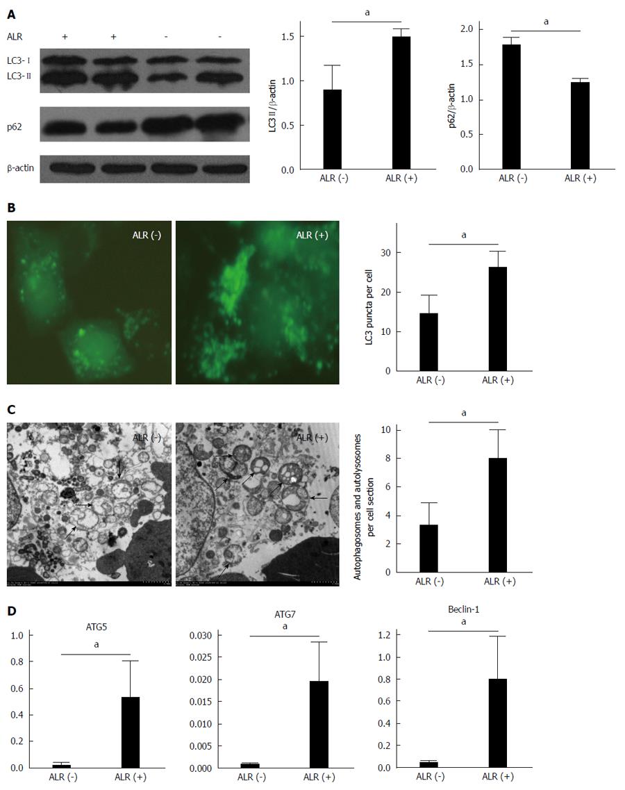
Figure 2 Augmenter of liver regeneration increases autophagic activity in HepG2 cells.
HepG2 cells were transfected with an augmenter of liver regeneration (ALR)-expressing plasmid or a control vector and cultured in Dulbecco’s modified Eagle’s medium for 6 h, followed by treatment with serum-free medium. Autophagy was observed at 24 h after serum deprivation. A: Total cell lysates were subjected to Western blot analysis with anti-LC3, anti-p62 and anti-β-actin antibodies. Densitometry was performed using ImageJ software to analyze the expression of LC3-II; B: GFP-LC3 puncta were determined by counting a total of more than 30 cells in triplicate samples; C: Digital images were obtained using an electron microscope (H-7650, Japan). The numbers of typical autophagosomes and autolysosomes in each cell section were counted randomly for more than 30 cells; D: The expression of ATG5, ATG7 and Beclin-1 mRNA was determined by qPCR. Digital data are presented as the mean ± SD from at least three independent experiments. aP < 0.05 ALR (-) vs ALR (+).
- Citation: Shi HB, Sun HQ, Shi HL, Ren F, Chen Y, Chen DX, Lou JL, Duan ZP. Autophagy in anti-apoptotic effect of augmenter of liver regeneration in HepG2 cells. World J Gastroenterol 2015; 21(17): 5250-5258
- URL: https://www.wjgnet.com/1007-9327/full/v21/i17/5250.htm
- DOI: https://dx.doi.org/10.3748/wjg.v21.i17.5250


