Copyright
©The Author(s) 2015.
World J Gastroenterol. May 7, 2015; 21(17): 5250-5258
Published online May 7, 2015. doi: 10.3748/wjg.v21.i17.5250
Published online May 7, 2015. doi: 10.3748/wjg.v21.i17.5250
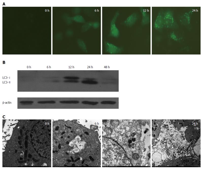
Figure 1 Starvation induces autophagic activation in HepG2 cells.
Autophagy in HepG2 cells was observed at 0, 12, 24, and 48 h after starvation caused by serum deprivation. A: HepG2 cells were transfected with the GFP-LC3 plasmid and cultured in Dulbecco’s modified Eagle’s medium for 6 h, followed by treatment with serum-free medium. Green puncta were observed using an inverted fluorescence microscope (Nikon Eclipse E800); B: Total cell lysates were subjected to Western blot analysis with anti-LC3 and anti-β-actin antibodies; C: Typical autophagosomes and autolysosomes (arrow) in HepG2 cells were observed via electron microscopy (H-7650, Japan).
- Citation: Shi HB, Sun HQ, Shi HL, Ren F, Chen Y, Chen DX, Lou JL, Duan ZP. Autophagy in anti-apoptotic effect of augmenter of liver regeneration in HepG2 cells. World J Gastroenterol 2015; 21(17): 5250-5258
- URL: https://www.wjgnet.com/1007-9327/full/v21/i17/5250.htm
- DOI: https://dx.doi.org/10.3748/wjg.v21.i17.5250


