Copyright
©The Author(s) 2015.
World J Gastroenterol. May 7, 2015; 21(17): 5231-5241
Published online May 7, 2015. doi: 10.3748/wjg.v21.i17.5231
Published online May 7, 2015. doi: 10.3748/wjg.v21.i17.5231
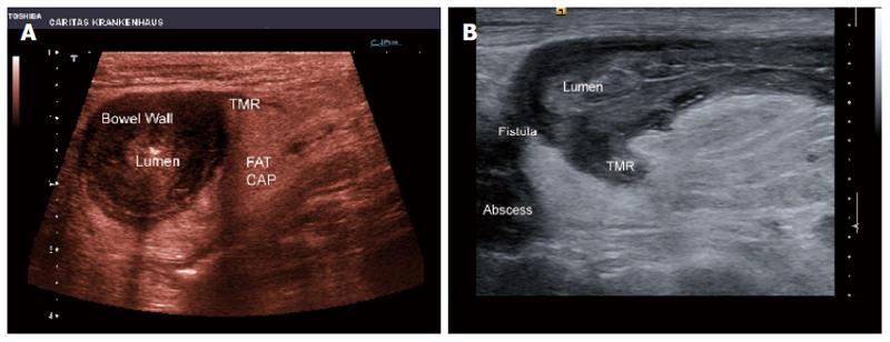
Figure 2 Segmental Crohn’s disease.
Note the “dirty” bowel wall thickening with loss of layer structure and the transmural inflammation without fistula and abscess formation. A: The periintestinal inflammatory reaction is shown by surrounding tissue (FAT CAP) (A). Crohn’s disease; B: Note the transmural inflammation (TMR) with fistula and abscess formation. The lumen is also indicated.
- Citation: Chiorean L, Schreiber-Dietrich D, Braden B, Cui XW, Buchhorn R, Chang JM, Dietrich CF. Ultrasonographic imaging of inflammatory bowel disease in pediatric patients. World J Gastroenterol 2015; 21(17): 5231-5241
- URL: https://www.wjgnet.com/1007-9327/full/v21/i17/5231.htm
- DOI: https://dx.doi.org/10.3748/wjg.v21.i17.5231


