Copyright
©The Author(s) 2015.
World J Gastroenterol. Apr 28, 2015; 21(16): 5105-5109
Published online Apr 28, 2015. doi: 10.3748/wjg.v21.i16.5105
Published online Apr 28, 2015. doi: 10.3748/wjg.v21.i16.5105
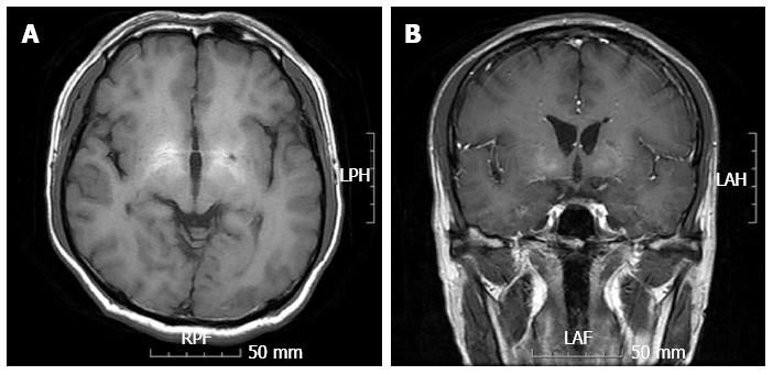
Figure 1 Magnetic resonance imaging.
Results of a magnetic resonance imaging brain scan exhibited symmetrical high-signal abnormalities in the basal ganglia on T1-weighted images, consistent with liver cirrhosis (A and B).
- Citation: Jo YM, Lee SW, Han SY, Baek YH, Ahn JH, Choi WJ, Lee JY, Kim SH, Yoon BA. Nonconvulsive status epilepticus disguising as hepatic encephalopathy. World J Gastroenterol 2015; 21(16): 5105-5109
- URL: https://www.wjgnet.com/1007-9327/full/v21/i16/5105.htm
- DOI: https://dx.doi.org/10.3748/wjg.v21.i16.5105


