Copyright
©The Author(s) 2015.
World J Gastroenterol. Apr 28, 2015; 21(16): 4946-4953
Published online Apr 28, 2015. doi: 10.3748/wjg.v21.i16.4946
Published online Apr 28, 2015. doi: 10.3748/wjg.v21.i16.4946
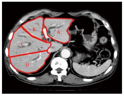
Figure 1 Computed tomography volumetric identification of liver sections.
Lateral section (A) and medial section (B) of the left liver, and anterior section (C) and posterior section (D) of the right liver.
- Citation: Takahashi E, Fukasawa M, Sato T, Takano S, Kadokura M, Shindo H, Yokota Y, Enomoto N. Biliary drainage strategy of unresectable malignant hilar strictures by computed tomography volumetry. World J Gastroenterol 2015; 21(16): 4946-4953
- URL: https://www.wjgnet.com/1007-9327/full/v21/i16/4946.htm
- DOI: https://dx.doi.org/10.3748/wjg.v21.i16.4946


