Copyright
©The Author(s) 2015.
World J Gastroenterol. Apr 28, 2015; 21(16): 4875-4882
Published online Apr 28, 2015. doi: 10.3748/wjg.v21.i16.4875
Published online Apr 28, 2015. doi: 10.3748/wjg.v21.i16.4875
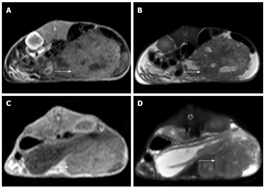
Figure 3 Magnetic resonance imaging of an implanted VX2 hepatocarcinoma with celiac implantation (shown with an arrow).
A, B: T1- and T2-weighted images of an implanted VX2 hepatocarcinoma with celiac implantation; C, D: T1- and T2-weighted images of another implanted VX2 hepatocarcinoma with celiac implantation.
- Citation: Chen Z, Kang Z, Xiao EH, Tong M, Xiao YD, Li HB. Comparison of two different laparotomy methods for modeling rabbit VX2 hepatocarcinoma. World J Gastroenterol 2015; 21(16): 4875-4882
- URL: https://www.wjgnet.com/1007-9327/full/v21/i16/4875.htm
- DOI: https://dx.doi.org/10.3748/wjg.v21.i16.4875


