Copyright
©The Author(s) 2015.
World J Gastroenterol. Apr 21, 2015; 21(15): 4607-4619
Published online Apr 21, 2015. doi: 10.3748/wjg.v21.i15.4607
Published online Apr 21, 2015. doi: 10.3748/wjg.v21.i15.4607
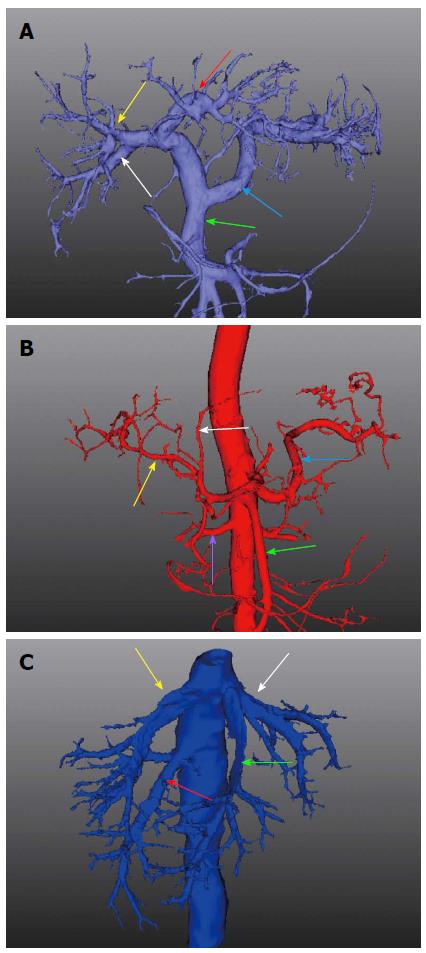
Figure 5 Three-dimensional images of the abdominal aorta system, portal vein system, and hepatic vein.
A: Three-dimensional (3D) image shows the portal vein system, which consists of the superior mesenteric vein (green arrow), splenic vein (blue arrow), left branch of the portal vein (red arrow), right anterior branch of the portal vein (yellow arrow), and right posterior branch of the portal vein (white arrow); B: 3D color-coded image of the abdominal aorta system: arteria hepatica propria including the left branch (white arrow) and right branch (yellow arrow), splenic artery (blue arrow), superior mesenteric artery (green arrow), and renal artery (purple arrow); C: Reconstruction of the outflow tract of the hepatic vein: left hepatic vein (white arrow), middle hepatic vein (green arrow), right hepatic vein (yellow arrow), and right posterior inferior hepatic vein (red arrow).
- Citation: Tian F, Wu JX, Rong WQ, Wang LM, Wu F, Yu WB, An SL, Liu FQ, Feng L, Bi C, Liu YH. Three-dimensional morphometric analysis for hepatectomy of centrally located hepatocellular carcinoma: A pilot study. World J Gastroenterol 2015; 21(15): 4607-4619
- URL: https://www.wjgnet.com/1007-9327/full/v21/i15/4607.htm
- DOI: https://dx.doi.org/10.3748/wjg.v21.i15.4607


