Copyright
©The Author(s) 2015.
World J Gastroenterol. Apr 14, 2015; 21(14): 4385-4390
Published online Apr 14, 2015. doi: 10.3748/wjg.v21.i14.4385
Published online Apr 14, 2015. doi: 10.3748/wjg.v21.i14.4385
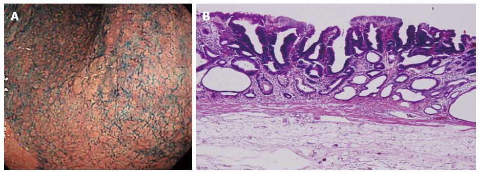
Figure 1 Initial early gastric cancer lesion in June 2005.
A: 0-IIa type well differentiated adenocarcinoma limited to the mucosa, 10 mm in size, without an ulcer scar, on the lesser curvature of the middle gastric body; B: Histopathological findings revealed a well differentiated mucosal adenocarcinoma, 10 mm in size, without lymphovascular involvement or ulcerative finding, as well as tumor-free margins.
- Citation: Shirahige A, Suzuki H, Oda I, Sekiguchi M, Mori G, Abe S, Nonaka S, Yoshinaga S, Sekine S, Kushima R, Saito Y, Fukagawa T, Katai H. Fatal submucosal invasive gastric adenosquamous carcinoma detected at surveillance after gastric endoscopic submucosal dissection. World J Gastroenterol 2015; 21(14): 4385-4390
- URL: https://www.wjgnet.com/1007-9327/full/v21/i14/4385.htm
- DOI: https://dx.doi.org/10.3748/wjg.v21.i14.4385


