Copyright
©The Author(s) 2015.
World J Gastroenterol. Apr 14, 2015; 21(14): 4275-4283
Published online Apr 14, 2015. doi: 10.3748/wjg.v21.i14.4275
Published online Apr 14, 2015. doi: 10.3748/wjg.v21.i14.4275
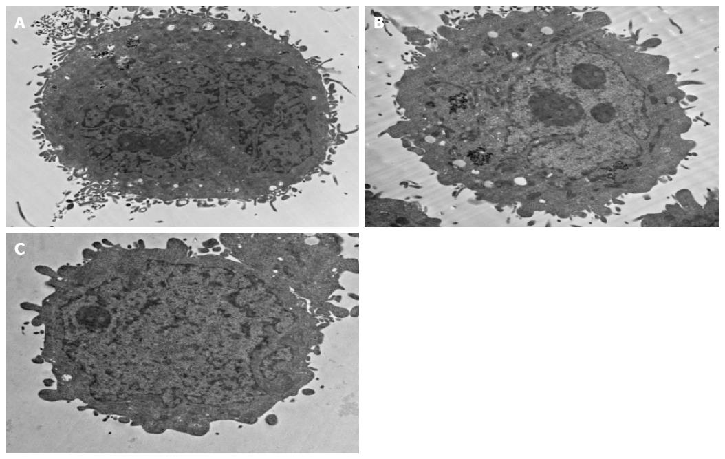
Figure 6 Transmission electron microscopy demonstrates the structure of the HepG2 cells under a magnification of × 10000.
A: HepG2 cell with expression of GPC3 receptors incubated with anti-GPC3-USPIONs probes, and the probes were seen on the surface of the cell and in the cell (black aggregation); B: HepG2 cell with expression of AFP receptors incubated with anti-AFP-USPIONs probes, and the probes were seen in the cell (black aggregation); C: HepG2 cell with expression of GPC3 receptors incubated with USPIONs probes, and no USPION was seen. USPION: Ultrasmall superparamagnetic iron oxide nanoparticle; AFP: α-fetoprotein; GPC3: Glypican 3.
- Citation: Li YW, Chen ZG, Zhao ZS, Li HL, Wang JC, Zhang ZM. Preparation of magnetic resonance probes using one-pot method for detection of hepatocellular carcinoma. World J Gastroenterol 2015; 21(14): 4275-4283
- URL: https://www.wjgnet.com/1007-9327/full/v21/i14/4275.htm
- DOI: https://dx.doi.org/10.3748/wjg.v21.i14.4275


