Copyright
©The Author(s) 2015.
World J Gastroenterol. Apr 14, 2015; 21(14): 4261-4267
Published online Apr 14, 2015. doi: 10.3748/wjg.v21.i14.4261
Published online Apr 14, 2015. doi: 10.3748/wjg.v21.i14.4261
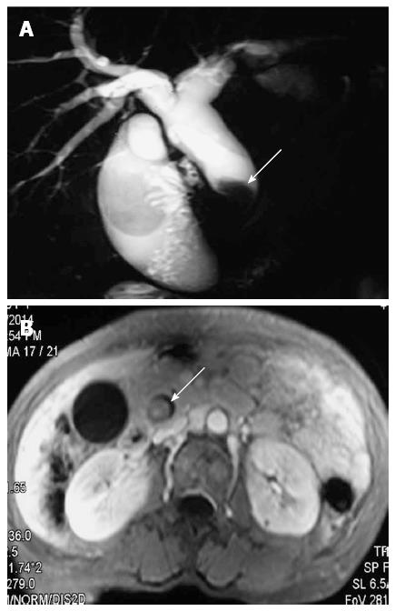
Figure 1 Imaging presentation of biliary tract intraductal papillary mucinous neoplasm.
A: Magnetic resonance cholangiography shows dilation of proximal biliary tract and a filling defect in the extrahepatic biliary tract (arrow); B: Magnetic resonance imaging shows an intraluminal polypoid lesion originating from the extrahepatic biliary tract (arrow).
- Citation: Wang X, Cai YQ, Chen YH, Liu XB. Biliary tract intraductal papillary mucinous neoplasm: Report of 19 cases. World J Gastroenterol 2015; 21(14): 4261-4267
- URL: https://www.wjgnet.com/1007-9327/full/v21/i14/4261.htm
- DOI: https://dx.doi.org/10.3748/wjg.v21.i14.4261


