Copyright
©The Author(s) 2015.
World J Gastroenterol. Apr 14, 2015; 21(14): 4184-4194
Published online Apr 14, 2015. doi: 10.3748/wjg.v21.i14.4184
Published online Apr 14, 2015. doi: 10.3748/wjg.v21.i14.4184
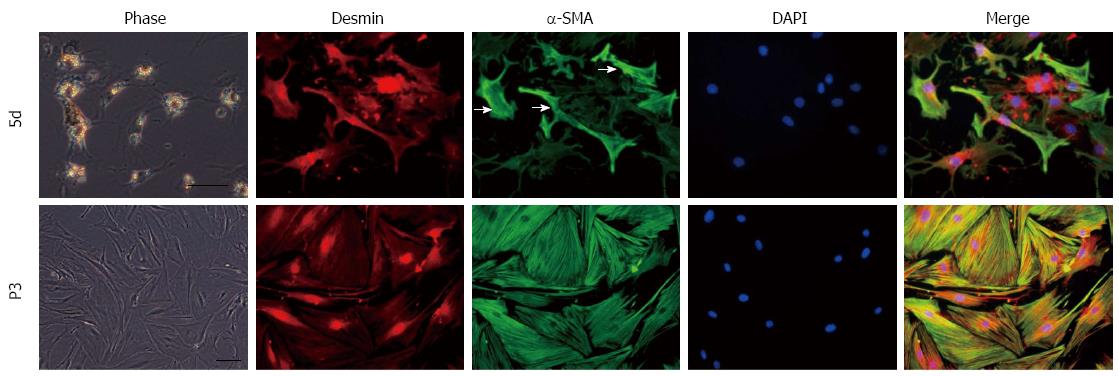
Figure 1 Morphological features and α-SMA expression of cultured hepatic stellate cells during the activation process.
Primary hepatic stellate cells (HSCs) were maintained on uncoated plastic dishes. HSCs presented a star-shaped appearance with fewer and smaller lipid droplets in the cytoplasm after primary culture for five days, and began to express α-SMA at non-uniform levels between cells. Subcultured HSCs evolved into myofibroblast-like cells without lipid droplets in the cytoplasm and expressed a uniform and higher level of α-SMA. Red: desmin; green: α-SMA; blue: nucleus (DAPI). Merged images are also shown. White arrows point to HSCs expressing high level of α-SMA. Bar = 100 μm.
- Citation: Chang WJ, Song LJ, Yi T, Shen KT, Wang HS, Gao XD, Li M, Xu JM, Niu WX, Qin XY. Early activated hepatic stellate cell-derived molecules reverse acute hepatic injury. World J Gastroenterol 2015; 21(14): 4184-4194
- URL: https://www.wjgnet.com/1007-9327/full/v21/i14/4184.htm
- DOI: https://dx.doi.org/10.3748/wjg.v21.i14.4184


