Copyright
©The Author(s) 2015.
World J Gastroenterol. Apr 7, 2015; 21(13): 4089-4095
Published online Apr 7, 2015. doi: 10.3748/wjg.v21.i13.4089
Published online Apr 7, 2015. doi: 10.3748/wjg.v21.i13.4089
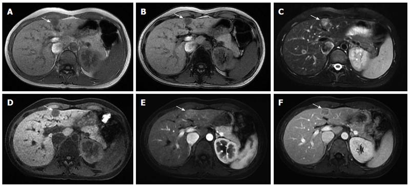
Figure 3 Liver magnetic resonance images demonstrated a small lymphoepithelioma-like cholangiocarcinoma at the medial segment of the liver (arrow).
A: In-phase T1-weighted image; B: Out of phase T1-weighted image; C: Axial T2-weighted image with fat suppression; D: Pre-contrast; E: Arterial T1-weighted image; F: Portal venous phase T1-weighted image with fat suppression.
- Citation: Liao TC, Liu CA, Chiu NC, Yeh YC, Chiou YY. Lymphoepithelioma-like cholangiocarcinoma: A mimic of hepatocellular carcinoma on imaging features. World J Gastroenterol 2015; 21(13): 4089-4095
- URL: https://www.wjgnet.com/1007-9327/full/v21/i13/4089.htm
- DOI: https://dx.doi.org/10.3748/wjg.v21.i13.4089


