Copyright
©The Author(s) 2015.
World J Gastroenterol. Mar 28, 2015; 21(12): 3759-3762
Published online Mar 28, 2015. doi: 10.3748/wjg.v21.i12.3759
Published online Mar 28, 2015. doi: 10.3748/wjg.v21.i12.3759
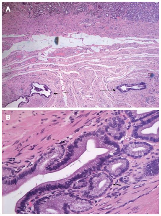
Figure 4 Histology.
Hematoxylin and eosin staining of nodular lesion specimens showed dilated cystic glands (arrows) in the A: Muscularis mucosa (× 50 magnification); B: Submucosal layers (× 400 magnification).
- Citation: Yu XF, Guo LW, Chen ST, Teng LS. Gastritis cystica profunda in a previously unoperated stomach: A case report. World J Gastroenterol 2015; 21(12): 3759-3762
- URL: https://www.wjgnet.com/1007-9327/full/v21/i12/3759.htm
- DOI: https://dx.doi.org/10.3748/wjg.v21.i12.3759


