Copyright
©The Author(s) 2015.
World J Gastroenterol. Mar 28, 2015; 21(12): 3579-3586
Published online Mar 28, 2015. doi: 10.3748/wjg.v21.i12.3579
Published online Mar 28, 2015. doi: 10.3748/wjg.v21.i12.3579
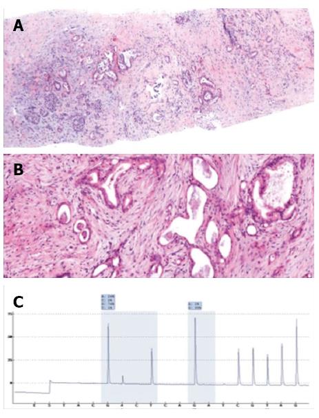
Figure 7 Pathologic and molecular analysis of a biopsied specimen.
A: Panoramic view (× 10) and B: High-power field (× 40) of a pancreatic core biopsy with hematoxylin and eosin staining showing atypical and irregularly displayed ductal structures in a desmoplastic stroma compatible with the diagnosis of ductal adenocarcinoma of the pancreas; C: Pyrogram demonstrating a mutation in codon 12.
- Citation: Tyng CJ, Almeida MFA, Barbosa PN, Bitencourt AG, Berg JAA, Maciel MS, Coimbra FJ, Schiavon LHO, Begnami MD, Guimarães MD, Zurstrassen CE, Chojniak R. Computed tomography-guided percutaneous core needle biopsy in pancreatic tumor diagnosis. World J Gastroenterol 2015; 21(12): 3579-3586
- URL: https://www.wjgnet.com/1007-9327/full/v21/i12/3579.htm
- DOI: https://dx.doi.org/10.3748/wjg.v21.i12.3579


