Copyright
©The Author(s) 2015.
World J Gastroenterol. Mar 28, 2015; 21(12): 3579-3586
Published online Mar 28, 2015. doi: 10.3748/wjg.v21.i12.3579
Published online Mar 28, 2015. doi: 10.3748/wjg.v21.i12.3579
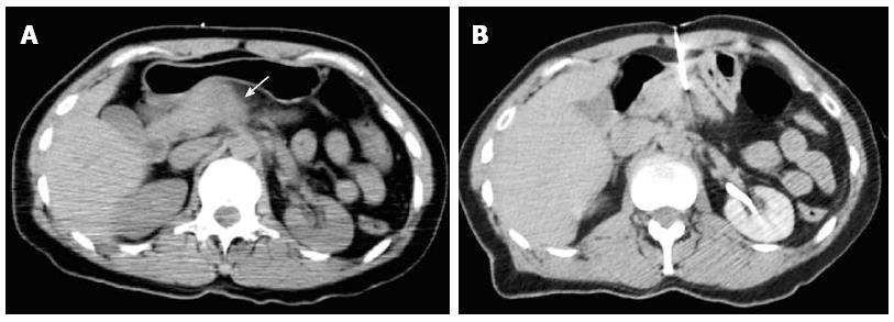
Figure 5 Percutaneous computed tomography-guided core needle biopsy of a pancreatic lesion using transgastric access.
A: Non-enhanced computed tomography showing a poorly defined nodule in the body of the pancreas (arrow). A posterior approach was considered difficult because of the interposition of large vessels in the needle path in the prone position, therefore, an anterior transgastric approach was used; B: Biopsy needle (20 G) placed adjacent to the pancreatic lesion through the stomach.
- Citation: Tyng CJ, Almeida MFA, Barbosa PN, Bitencourt AG, Berg JAA, Maciel MS, Coimbra FJ, Schiavon LHO, Begnami MD, Guimarães MD, Zurstrassen CE, Chojniak R. Computed tomography-guided percutaneous core needle biopsy in pancreatic tumor diagnosis. World J Gastroenterol 2015; 21(12): 3579-3586
- URL: https://www.wjgnet.com/1007-9327/full/v21/i12/3579.htm
- DOI: https://dx.doi.org/10.3748/wjg.v21.i12.3579


