Copyright
©The Author(s) 2015.
World J Gastroenterol. Mar 21, 2015; 21(11): 3402-3408
Published online Mar 21, 2015. doi: 10.3748/wjg.v21.i11.3402
Published online Mar 21, 2015. doi: 10.3748/wjg.v21.i11.3402
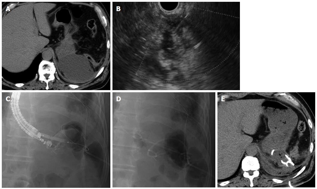
Figure 4 Case 4.
A: Computed tomography (CT) reveals fluid collection at the dorsal side of the fornix close to the gastric wall; B: Endoscopic ultrasound (EUS) showing the fluid collection, which was punctured by a 19-gauge needle; C and D: Fluoroscopy image showing placement of the percutaneous drain and insertion of the 6-mm balloon catheter over the guidewire. Both a 7-Fr double pigtail stent and a 6-Fr nasal biliary catheter are placed; E: Ten days after EUS-guided drainage, CT reveals that the fluid collection decreased.
- Citation: Mandai K, Uno K, Yasuda K. Endoscopic ultrasound-guided drainage of postoperative intra-abdominal abscesses. World J Gastroenterol 2015; 21(11): 3402-3408
- URL: https://www.wjgnet.com/1007-9327/full/v21/i11/3402.htm
- DOI: https://dx.doi.org/10.3748/wjg.v21.i11.3402


