Copyright
©The Author(s) 2015.
World J Gastroenterol. Mar 21, 2015; 21(11): 3380-3387
Published online Mar 21, 2015. doi: 10.3748/wjg.v21.i11.3380
Published online Mar 21, 2015. doi: 10.3748/wjg.v21.i11.3380
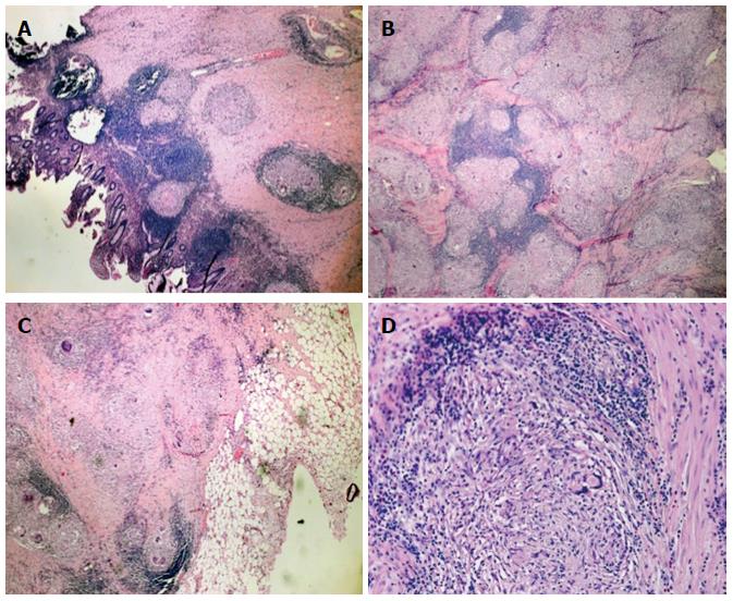
Figure 4 Histologic examination of the intraoperative specimen.
Hematoxylin and eosin staining showed A: Noncaseating epithelioid granulomas in the colonic wall (magnification × 5); B: Confluent granulomata in the colonic wall (magnification × 10); C: Perivisceral involvement (magnification × 5); D: Microscopic aspects of the sarcoidotic granulomas (magnification × 20).
- Citation: Erra P, Crusco S, Nugnes L, Pollio AM, Di Pilla G, Biondi G, Vigliardi G. Colonic sarcoidosis: Unusual onset of a systemic disease. World J Gastroenterol 2015; 21(11): 3380-3387
- URL: https://www.wjgnet.com/1007-9327/full/v21/i11/3380.htm
- DOI: https://dx.doi.org/10.3748/wjg.v21.i11.3380


