Copyright
©The Author(s) 2015.
World J Gastroenterol. Mar 21, 2015; 21(11): 3380-3387
Published online Mar 21, 2015. doi: 10.3748/wjg.v21.i11.3380
Published online Mar 21, 2015. doi: 10.3748/wjg.v21.i11.3380
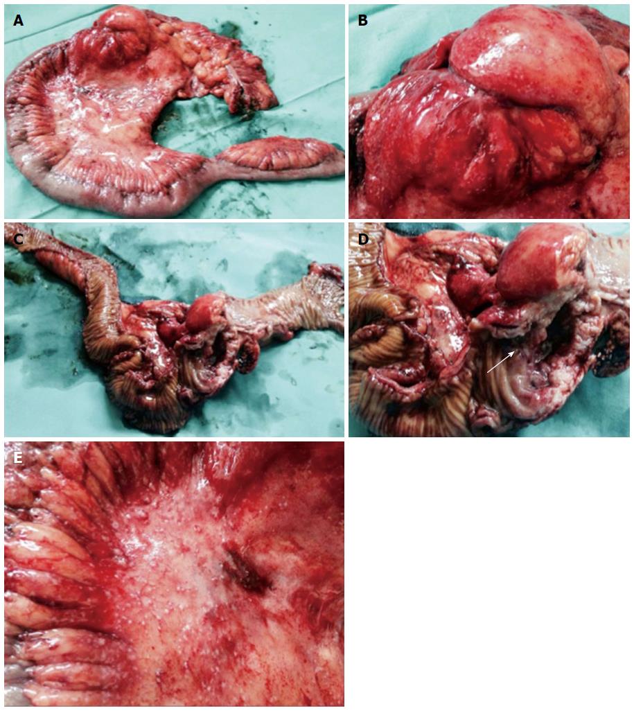
Figure 3 Resected specimen after laparotomy.
A: Right hemicolectomy was performed; B-D: A voluminous stenotic ulcerated lesion of the colonic wall in proximity to the ileocecal valve (arrow in D) was observed; E: Numerous peritoneal micronodules near the colonic lesion are shown.
- Citation: Erra P, Crusco S, Nugnes L, Pollio AM, Di Pilla G, Biondi G, Vigliardi G. Colonic sarcoidosis: Unusual onset of a systemic disease. World J Gastroenterol 2015; 21(11): 3380-3387
- URL: https://www.wjgnet.com/1007-9327/full/v21/i11/3380.htm
- DOI: https://dx.doi.org/10.3748/wjg.v21.i11.3380


