Copyright
©The Author(s) 2015.
World J Gastroenterol. Mar 14, 2015; 21(10): 3121-3126
Published online Mar 14, 2015. doi: 10.3748/wjg.v21.i10.3121
Published online Mar 14, 2015. doi: 10.3748/wjg.v21.i10.3121
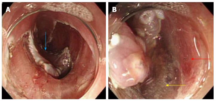
Figure 2 Endoscopic submucosal dissection for esophageal cancer.
A: Depressed area at the center of the lesion became noticeable during the course of submucosal dissection (blue arrow); B: Muscularis propria layer abruptly ended at the center of the cancer lesion. The red arrow indicates the muscularis propria layer, and the yellow arrow indicates the area of the muscularis propria layer defect.
- Citation: Tanaka S, Toyonaga T, Ohara Y, Yoshizaki T, Kawara F, Ishida T, Hoshi N, Morita Y, Azuma T. Esophageal diverticulum exposed during endoscopic submucosal dissection of superficial cancer. World J Gastroenterol 2015; 21(10): 3121-3126
- URL: https://www.wjgnet.com/1007-9327/full/v21/i10/3121.htm
- DOI: https://dx.doi.org/10.3748/wjg.v21.i10.3121


