Copyright
©The Author(s) 2015.
World J Gastroenterol. Mar 14, 2015; 21(10): 3121-3126
Published online Mar 14, 2015. doi: 10.3748/wjg.v21.i10.3121
Published online Mar 14, 2015. doi: 10.3748/wjg.v21.i10.3121
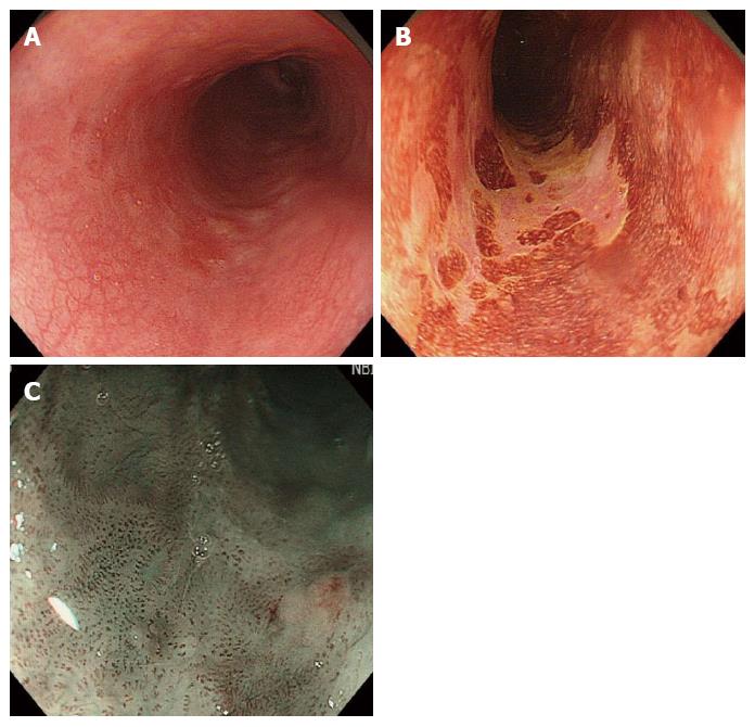
Figure 1 Endoscopic observation of esophageal cancer.
A, B: Lesion had expanded to half of the circumference of the lumen; C: In magnified narrow band imaging (NBI) observation, intraepithelial papillary capillary loop (IPCL) was detected as a small dot pattern.
- Citation: Tanaka S, Toyonaga T, Ohara Y, Yoshizaki T, Kawara F, Ishida T, Hoshi N, Morita Y, Azuma T. Esophageal diverticulum exposed during endoscopic submucosal dissection of superficial cancer. World J Gastroenterol 2015; 21(10): 3121-3126
- URL: https://www.wjgnet.com/1007-9327/full/v21/i10/3121.htm
- DOI: https://dx.doi.org/10.3748/wjg.v21.i10.3121


