Copyright
©The Author(s) 2015.
World J Gastroenterol. Mar 14, 2015; 21(10): 3035-3040
Published online Mar 14, 2015. doi: 10.3748/wjg.v21.i10.3035
Published online Mar 14, 2015. doi: 10.3748/wjg.v21.i10.3035
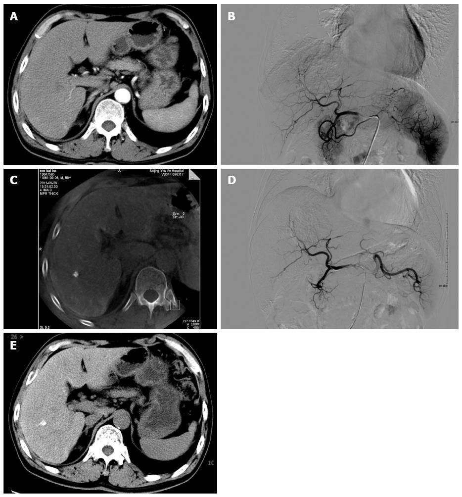
Figure 1 Imaging findings in a 50-year-old man diagnosed with hepatocellular carcinoma.
Re-examination after transcatheter arterial chemoembolization (TACE). A: No clear lesion was found on pre-procedure 64-slice CT; B: No clear tumor staining was found following digital subtraction angiography examination; C: C-arm Lipiodol computed tomography (CT) scan after diagnostic embolization. A new Lipiodol lesion was found in the 6th liver segment; D: Gelfoam particles were injected into the tumor-nourishing blood vessel; E: New hepatocellular carcinoma lesion in the 6th segment was confirmed by CT examination performed 12 d after TACE.
- Citation: Li JJ, Zheng JS, Cui SC, Cui XW, Hu CX, Fang D, Ye LC. C-arm Lipiodol CT in transcatheter arterial chemoembolization for small hepatocellular carcinoma. World J Gastroenterol 2015; 21(10): 3035-3040
- URL: https://www.wjgnet.com/1007-9327/full/v21/i10/3035.htm
- DOI: https://dx.doi.org/10.3748/wjg.v21.i10.3035


