Copyright
©The Author(s) 2015.
World J Gastroenterol. Mar 14, 2015; 21(10): 2997-3004
Published online Mar 14, 2015. doi: 10.3748/wjg.v21.i10.2997
Published online Mar 14, 2015. doi: 10.3748/wjg.v21.i10.2997
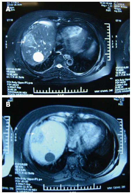
Figure 1 Magnetic resonance imaging of the liver in a patient with medium-sized hepatocellular carcinoma before and after treatment with microwave ablation.
A: Magnetic resonance imaging of liver before treatment with microwave ablation (MWA). Medium-sized hepatocellular carcinoma (HCC) is labeled (arrow); B: MRI of liver at 1 mo after treatment with MWA. Medium-sized HCC is labeled (arrow). The tumor diameter was 4 cm × 4 cm. The power was set to 100 W. The ablation time was 3 min.
- Citation: Sun AX, Cheng ZL, Wu PP, Sheng YH, Qu XJ, Lu W, Zhao CG, Qian GJ. Clinical outcome of medium-sized hepatocellular carcinoma treated with microwave ablation. World J Gastroenterol 2015; 21(10): 2997-3004
- URL: https://www.wjgnet.com/1007-9327/full/v21/i10/2997.htm
- DOI: https://dx.doi.org/10.3748/wjg.v21.i10.2997


