Copyright
©The Author(s) 2015.
World J Gastroenterol. Jan 7, 2015; 21(1): 60-83
Published online Jan 7, 2015. doi: 10.3748/wjg.v21.i1.60
Published online Jan 7, 2015. doi: 10.3748/wjg.v21.i1.60
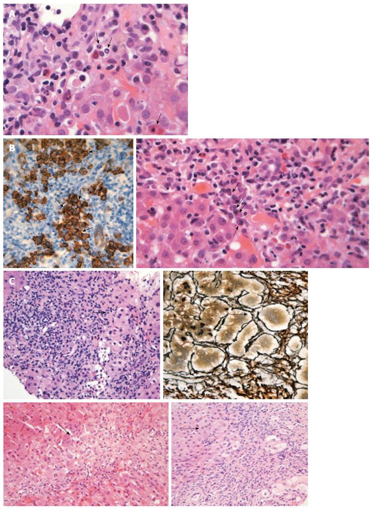
Figure 3 Liver histology in autoimmune hepatitis.
A: Interface hepatitis with genuine apoptotic bodies (arrows) in a case with autoimmune hepatitis-type 1 (hematoxylin and eosin staining). See also the typical rim of condensed chromatin at the nuclear membrane and ordinary acidophilic bodies, at various stages of development; B: Intense portal plasmacytosis in autoimmune hepatitis (arrows in the right panel; hematoxylin and eosin staining). The plasma cells are in clusters, better seen after immunostaining with CD138 (arrows in dark brown staining of the left panel). The intensity of plasmacytosis can be useful in discriminating autoimmune hepatitis from viral hepatitis whereas there is also possible prognostic information as portal plasma cell infiltrates while on immunosuppression are associated with relapse upon drug cessation; C: Various examples of rosette formation (hematoxylin and eosin staining; periportal, periseptal in a previous collapse and with reticulin staining seen in the upper right panel; see arrows).
- Citation: Gatselis NK, Zachou K, Koukoulis GK, Dalekos GN. Autoimmune hepatitis, one disease with many faces: Etiopathogenetic, clinico-laboratory and histological characteristics. World J Gastroenterol 2015; 21(1): 60-83
- URL: https://www.wjgnet.com/1007-9327/full/v21/i1/60.htm
- DOI: https://dx.doi.org/10.3748/wjg.v21.i1.60


