Copyright
©The Author(s) 2015.
World J Gastroenterol. Jan 7, 2015; 21(1): 369-372
Published online Jan 7, 2015. doi: 10.3748/wjg.v21.i1.369
Published online Jan 7, 2015. doi: 10.3748/wjg.v21.i1.369
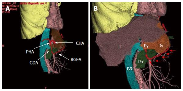
Figure 2 Three-dimensional computed tomography.
A: The courses of the branches of celiac artery were well visualized while obtaining surgical navigation; B: Relationship with the gastric tube and surrounding structures were well visualized while obtaining surgical navigation. Arrows shows the running of right gastroepiploic artery. L: Liver, IVC: Inferior vena cava; Pa: pancreas; G: Gastric tube; Py: Pylorus; CHA: Common hepatic artery; RGEA: Right gastroepiploic artery; GDA: Gastroduodenal artery; PHA: Proper hepatic artery.
- Citation: Nakano T, Sakurai T, Maruyama S, Ozawa Y, Kamei T, Miyata G, Ohuchi N. Indocyanine green fluorescence and three-dimensional imaging of right gastroepiploic artery in gastric tube cancer. World J Gastroenterol 2015; 21(1): 369-372
- URL: https://www.wjgnet.com/1007-9327/full/v21/i1/369.htm
- DOI: https://dx.doi.org/10.3748/wjg.v21.i1.369


