Copyright
©The Author(s) 2015.
World J Gastroenterol. Jan 7, 2015; 21(1): 187-195
Published online Jan 7, 2015. doi: 10.3748/wjg.v21.i1.187
Published online Jan 7, 2015. doi: 10.3748/wjg.v21.i1.187
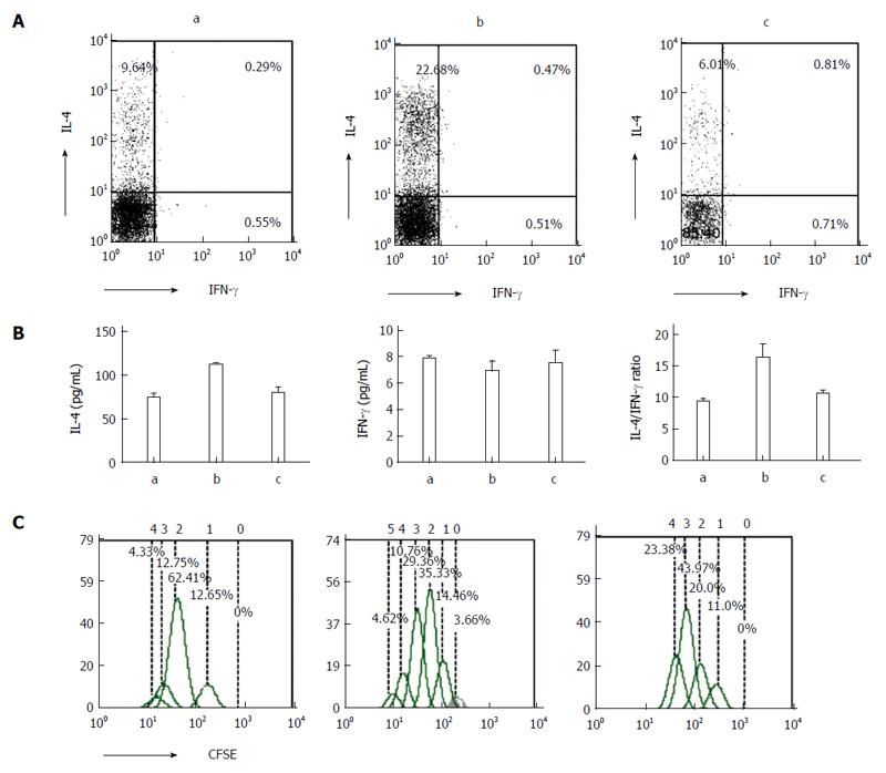
Figure 5 T lymphocyte differentiation and proliferation induced by mouse intestinal epithelial cells.
A: Flow cytometric analysis of interferon (IFN)-γ and interleukin (IL)-4 levels in CD4+ T cells; B: Enzyme-linked immunosorbent assay of IFN-γ and IL-4 levels in co-culture supernatants; C: Flow cytometric analysis of T cell proliferation (0, undivided cells; 1, generation 1; 2, generation 2; 3, generation 3; 4, generation 4; 5, generation 5). a: Control group, b: Dextran sodium sulfate-treated group, c: Anti-P-selectin lectin-EGF domain monoclonal antibody-treated group; aP < 0.05 vs controls; cP < 0.05 vs dextran sodium sulfate-treated group. No significant changes in IFN-γ groups.
- Citation: Zeng JQ, Xu CD, Zhou T, Wu J, Lin K, Liu W, Wang XQ. Enterocyte dendritic cell-specific intercellular adhesion molecule-3-grabbing non-integrin expression in inflammatory bowel disease. World J Gastroenterol 2015; 21(1): 187-195
- URL: https://www.wjgnet.com/1007-9327/full/v21/i1/187.htm
- DOI: https://dx.doi.org/10.3748/wjg.v21.i1.187


