Copyright
©2014 Baishideng Publishing Group Co.
World J Gastroenterol. Feb 28, 2014; 20(8): 2051-2061
Published online Feb 28, 2014. doi: 10.3748/wjg.v20.i8.2051
Published online Feb 28, 2014. doi: 10.3748/wjg.v20.i8.2051
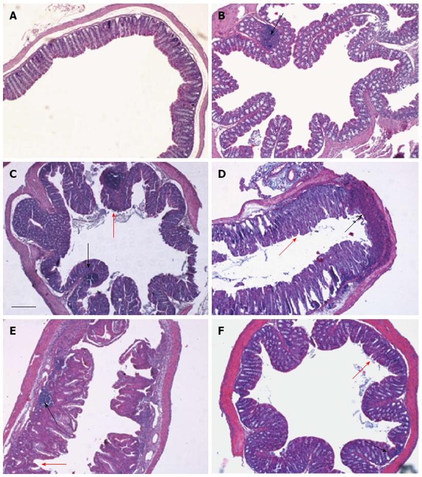
Figure 4 Differences in histological parameters during experimental colitis.
Colons were collected from DSS-treated mice on days 3 (B), 7 (C) 13 (D), 19 (E) and 29 (F). In comparison to control mice (A), histopathological changes in individual crypts are shown in representative hematoxylin and eosin-stained sections. Loss of crypt architecture associated with epithelial damage and flattened villi (red arrows) and leukocyte infiltration (black arrows) are evident following DSS treatment (bar = 200 μm).
- Citation: Fazio LD, Cavazza E, Spisni E, Strillacci A, Centanni M, Candela M, Praticò C, Campieri M, Ricci C, Valerii MC. Longitudinal analysis of inflammation and microbiota dynamics in a model of mild chronic dextran sulfate sodium-induced colitis in mice. World J Gastroenterol 2014; 20(8): 2051-2061
- URL: https://www.wjgnet.com/1007-9327/full/v20/i8/2051.htm
- DOI: https://dx.doi.org/10.3748/wjg.v20.i8.2051


