Copyright
©2014 Baishideng Publishing Group Co.
World J Gastroenterol. Feb 28, 2014; 20(8): 2030-2041
Published online Feb 28, 2014. doi: 10.3748/wjg.v20.i8.2030
Published online Feb 28, 2014. doi: 10.3748/wjg.v20.i8.2030
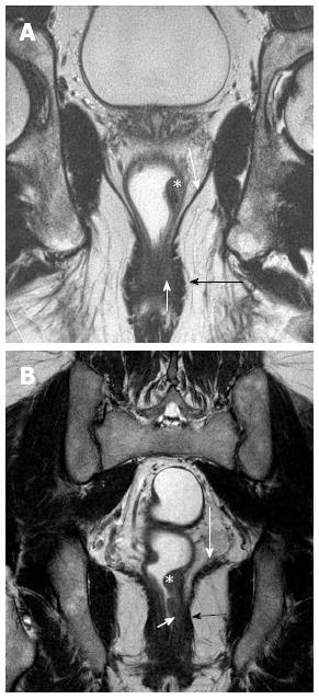
Figure 10 Coronal section T2 weighted magnetic resonance imaging to see level of tumor for planning surgery.
A and B show tumor (asterisk) along the left lateral wall, that reaches up to the internal sphincter (short white arrows) in B, but spares it in A. The uninvolved external sphincter (darkly hypointense) is shown by black arrows. The long white arrows in A and B shows the spared levator ani.
- Citation: Saklani AP, Bae SU, Clayton A, Kim NK. Magnetic resonance imaging in rectal cancer: A surgeon’s perspective. World J Gastroenterol 2014; 20(8): 2030-2041
- URL: https://www.wjgnet.com/1007-9327/full/v20/i8/2030.htm
- DOI: https://dx.doi.org/10.3748/wjg.v20.i8.2030


