Copyright
©2014 Baishideng Publishing Group Co.
World J Gastroenterol. Feb 28, 2014; 20(8): 2014-2022
Published online Feb 28, 2014. doi: 10.3748/wjg.v20.i8.2014
Published online Feb 28, 2014. doi: 10.3748/wjg.v20.i8.2014
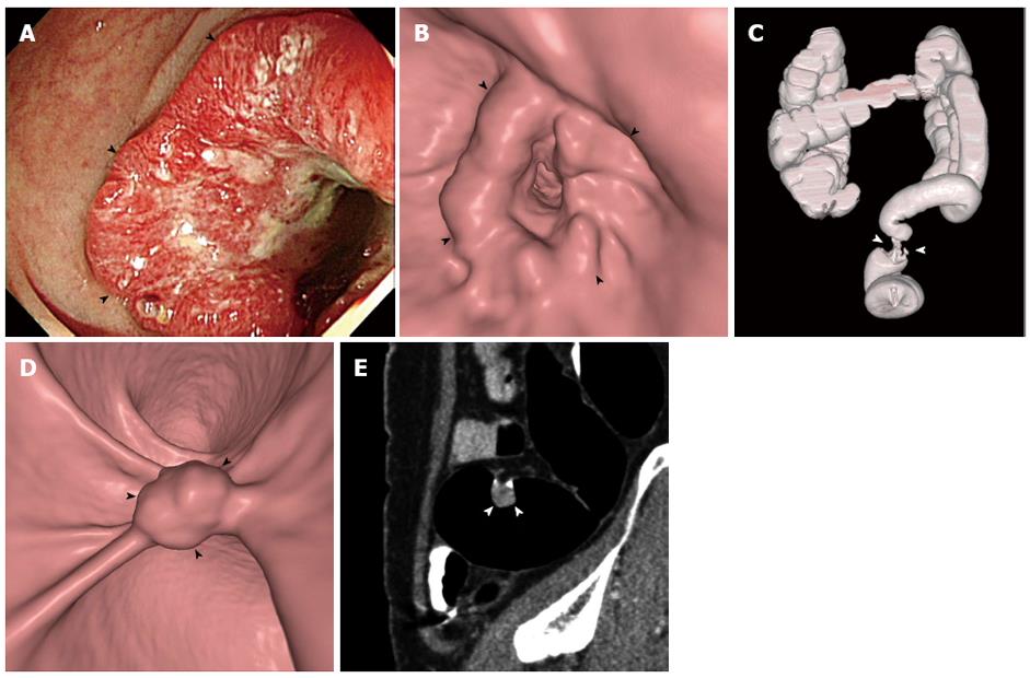
Figure 1 A 55-year-old woman with occlusive cancer in the upper rectum and a 17-mm synchronous cancer in the sigmoid colon.
A-C: Colonoscopic (A), three-dimensional endoluminal computed tomographic (CT) colonographic (i.e., virtual colonoscopic) (B), and three-dimensional volume-rendered (C) images of the colon show a luminal-encircling occlusive mass (arrowheads) in the upper rectum that impeded the passage of a colonoscope; D, E: Three-dimensional endoluminal (D) and two-dimensional sagittal (E) CT colonographic images show a 17-mm polyp (arrowheads) unassociated with invasive features in the sigmoid colon. The lesion was removed by surgery and pathologically confirmed as a cancer confined to the mucosa.
- Citation: Hong N, Park SH. CT colonography in the diagnosis and management of colorectal cancer: Emphasis on pre- and post-surgical evaluation. World J Gastroenterol 2014; 20(8): 2014-2022
- URL: https://www.wjgnet.com/1007-9327/full/v20/i8/2014.htm
- DOI: https://dx.doi.org/10.3748/wjg.v20.i8.2014


