Copyright
©2014 Baishideng Publishing Group Co.
World J Gastroenterol. Feb 21, 2014; 20(7): 1807-1821
Published online Feb 21, 2014. doi: 10.3748/wjg.v20.i7.1807
Published online Feb 21, 2014. doi: 10.3748/wjg.v20.i7.1807
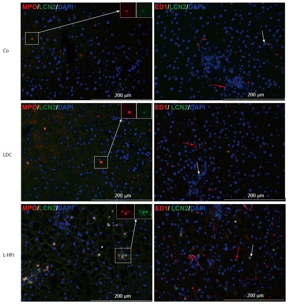
Figure 7 Representative photomicrographs show immunolocalization of key markers of Neutrophil granulocytes infiltration.
Myeloperoxidase (MPO) double-stained with LCN-2 (left-panel), and tissue macrophages (ED1) plus LCN-2 (right-panel) at week 4; A co-localization of LCN-2 and MPO can be detected in liver sections. The black cavities (vesicles) that were marked by Star in L-HFr micrographs resulted from washing of fat from hepatocytes during fixation step. Red arrows in right panel show ED1+ cells. Scale bar = 200 μm. Co: Chow diet, LDC: Lieber-DeCarli liquid diet; L-HFr: LDC + 70% fructose.
- Citation: Alwahsh SM, Xu M, Seyhan HA, Ahmad S, Mihm S, Ramadori G, Schultze FC. Diet high in fructose leads to an overexpression of lipocalin-2 in rat fatty liver. World J Gastroenterol 2014; 20(7): 1807-1821
- URL: https://www.wjgnet.com/1007-9327/full/v20/i7/1807.htm
- DOI: https://dx.doi.org/10.3748/wjg.v20.i7.1807


