Copyright
©2014 Baishideng Publishing Group Co.
World J Gastroenterol. Feb 21, 2014; 20(7): 1797-1806
Published online Feb 21, 2014. doi: 10.3748/wjg.v20.i7.1797
Published online Feb 21, 2014. doi: 10.3748/wjg.v20.i7.1797
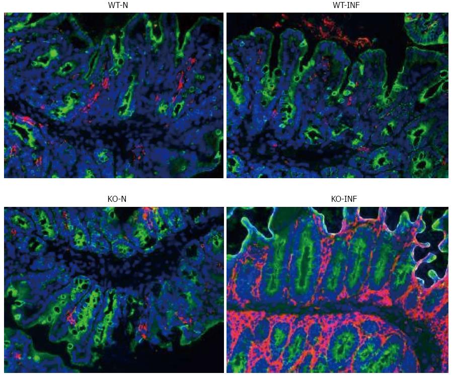
Figure 7 Dendritic cell infiltration associated with Trichuris muris infection in the proximal colon of FVB (WT) and mdr1a-/- (PGP-KO) mice.
Fluorescence staining (pink) for dendritic cells in frozen sections of proximal colon from naïve (WT-N; KO-N) and Trichuris muris (T. muris) infected (WT-INF; KO-INF) mice. Tissues were removed on day 19 post infection in T. muris infected mice. Original magnification × 20. Images are representative of tissues from 5 mice in each group.
-
Citation: Bhardwaj EK, Else KJ, Rogan MT, Warhurst G. Increased susceptibility to
Trichuris muris infection and exacerbation of colitis in Mdr1a-/- mice. World J Gastroenterol 2014; 20(7): 1797-1806 - URL: https://www.wjgnet.com/1007-9327/full/v20/i7/1797.htm
- DOI: https://dx.doi.org/10.3748/wjg.v20.i7.1797


